Local neurotrophin effects on central trigeminal axon growth patterns
- PMID: 15246692
- PMCID: PMC4283502
- DOI: 10.1016/j.devbrainres.2004.03.017
Local neurotrophin effects on central trigeminal axon growth patterns
Abstract
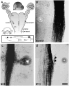
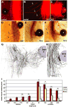
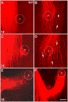
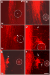
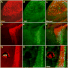
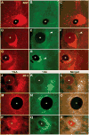
In dissociated cell and wholemount explant cultures of the embryonic trigeminal pathway NGF promotes exuberant elongation of trigeminal ganglion (TG) axons, whereas NT-3 leads to precocious arborization [J. Comp. Neurol. 425 (2000) 202]. In the present study, we investigated the axonal effects of local applications of NGF and NT-3. We placed small sepharose beads loaded with either NGF or NT-3 along the lateral edge of the central trigeminal tract in TG-brainstem intact wholemount explant cultures prepared from embryonic day 15 rats. Labeling of the TG with carbocyanine dye, DiI, revealed that NGF induces local defasciculation and diversion of trigeminal axons. Numerous axons leave the tract, grow towards the bead and engulf it, while some axons grow away from the neurotrophin source. NT-3, on the other hand, induced localized interstitial branching and formation of neuritic tangles in the vicinity of the neurotrophin source. Double immunocytochemistry showed that axons responding to NGF were predominantly TrkA-positive, whereas both TrkA and TrkC-positive axons responded to NT-3. Our results indicate that localized neurotrophin sources along the routes of embryonic sensory axons in the central nervous system, far away from their parent cell bodies, can alter restricted axonal pathways and induce elongation, arborization responses.
Figures
Similar articles
-
Morphometric analysis of embryonic rat trigeminal neurons treated with different neurotrophins.Anat Rec A Discov Mol Cell Evol Biol. 2004 Apr;277(2):396-407. doi: 10.1002/ar.a.20029. Anat Rec A Discov Mol Cell Evol Biol. 2004. PMID: 15052666 Free PMC article.
-
Regulation of neurotrophin-induced axonal responses via Rho GTPases.J Comp Neurol. 2001 Oct 1;438(4):377-87. doi: 10.1002/cne.1321. J Comp Neurol. 2001. PMID: 11559894 Free PMC article.
-
Differential effects of NGF and NT-3 on embryonic trigeminal axon growth patterns.J Comp Neurol. 2000 Sep 18;425(2):202-18. doi: 10.1002/1096-9861(20000918)425:2<202::aid-cne4>3.0.co;2-t. J Comp Neurol. 2000. PMID: 10954840 Free PMC article.
-
NGF synthesis and NGF receptor expression in the embryonic mouse trigeminal system.J Physiol (Paris). 1990;84(1):100-3. J Physiol (Paris). 1990. PMID: 2162955 Review.
-
Target attraction: are developing axons guided by chemotropism?Trends Neurosci. 1991 Jul;14(7):303-10. doi: 10.1016/0166-2236(91)90142-h. Trends Neurosci. 1991. PMID: 1719678 Review.
Cited by
-
Differential Trk expression in explant and dissociated trigeminal ganglion cell cultures.J Neurobiol. 2005 Aug;64(2):145-56. doi: 10.1002/neu.20134. J Neurobiol. 2005. PMID: 15828064 Free PMC article.
-
Null mutations of NT-3 and Bax affect trigeminal ganglion cell number but not brainstem barrelette pattern formation.Somatosens Mot Res. 2013 Sep;30(3):114-9. doi: 10.3109/08990220.2013.775118. Epub 2013 Apr 24. Somatosens Mot Res. 2013. PMID: 23614607 Free PMC article.
-
Molecular determinants of the face map development in the trigeminal brainstem.Anat Rec A Discov Mol Cell Evol Biol. 2006 Feb;288(2):121-34. doi: 10.1002/ar.a.20285. Anat Rec A Discov Mol Cell Evol Biol. 2006. PMID: 16432893 Free PMC article. Review.
-
SAD kinases sculpt axonal arbors of sensory neurons through long- and short-term responses to neurotrophin signals.Neuron. 2013 Jul 10;79(1):39-53. doi: 10.1016/j.neuron.2013.05.017. Epub 2013 Jun 20. Neuron. 2013. PMID: 23790753 Free PMC article.
-
Neocortical axon arbors trade-off material and conduction delay conservation.PLoS Comput Biol. 2010 Mar 12;6(3):e1000711. doi: 10.1371/journal.pcbi.1000711. PLoS Comput Biol. 2010. PMID: 20300651 Free PMC article.
References
-
- Buchman VL, Davies AM. Different neurotrophins are expressed and act in a developmental sequence to promote the survival of embryonic sensory neurons. Development. 1993;118:989–1001. - PubMed
-
- Campenot RB. NGF and the local control of nerve terminal growth. J Neurobiol. 1994;25:599–611. - PubMed
-
- Castellani V, Bolz J. Opposing roles for neurotrophin-3 in targeting and collateral formation of distinct sets of developing cortical neurons. Development. 1999;126:3335–3345. - PubMed
-
- Cohen-Corey S, Fraser SE. Effects of brain-derived neurotrophic factor on optic axon branching and remodeling in vivo. Nature. 1995;378:192–196. - PubMed
Publication types
MeSH terms
Substances
Grants and funding
LinkOut - more resources
Full Text Sources
Research Materials
Miscellaneous

