. 2004 Sep 24;279(39):40699-706.
doi: 10.1074/jbc.M402697200. Epub 2004 Jul 6.

Disruption of cortical actin in skeletal muscle demonstrates an essential role of the cytoskeleton in glucose transporter 4 translocation in insulin-sensitive tissues

Affiliations

Disruption of cortical actin in skeletal muscle demonstrates an essential role of the cytoskeleton in glucose transporter 4 translocation in insulin-sensitive tissues

Joseph T Brozinick Jr et al. J Biol Chem. .

Abstract

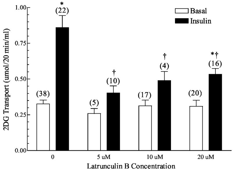
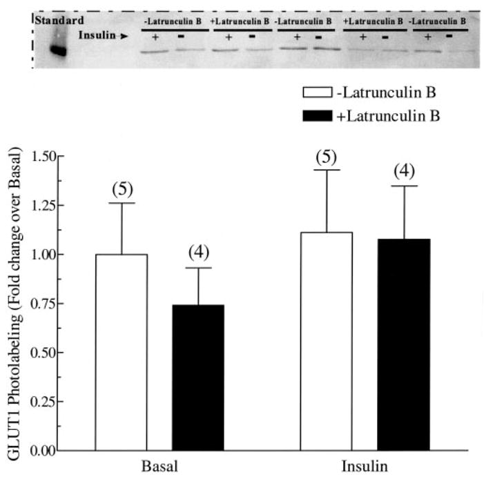
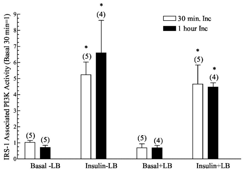
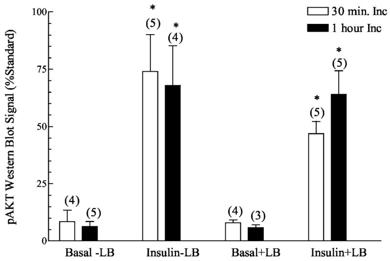
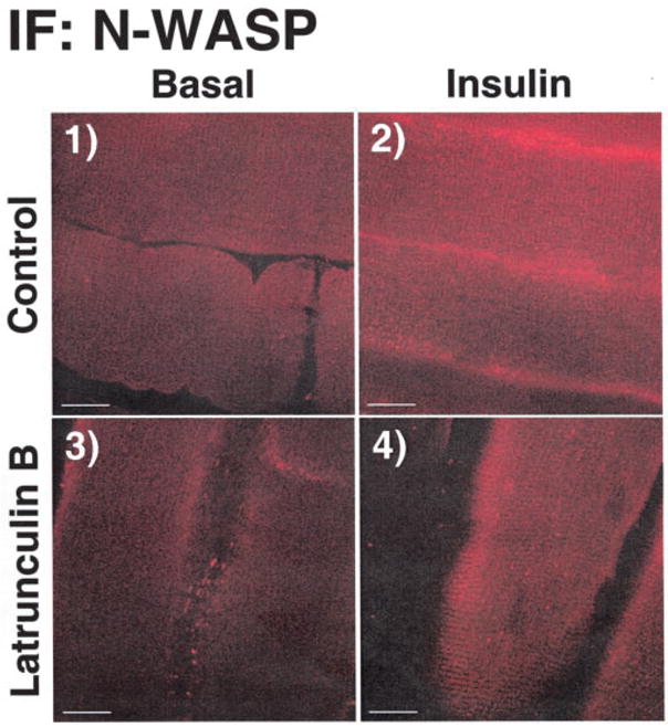
Cell culture work suggests that signaling to polymerize cortical filamentous actin (F-actin) represents a required pathway for the optimal redistribution of the insulin-responsive glucose transporter, GLUT4, to the plasma membrane. Recent in vitro study further suggests that the actin-regulatory neural Wiskott-Aldrich syndrome protein (N-WASP) mediates the effect of insulin on the actin filament network. Here we tested whether similar cytoskeletal mechanics are essential for insulin-regulated glucose transport in isolated rat epitrochlearis skeletal muscle. Microscopic analysis revealed that cortical F-actin is markedly diminished in muscle exposed to latrunculin B. Depolymerization of cortical F-actin with latrunculin B caused a time- and concentration-dependent decline in 2-deoxyglucose transport. The loss of cortical F-actin and glucose transport was paralleled by a decline in insulin-stimulated GLUT4 translocation, as assessed by photolabeling of cell surface GLUT4 with Bio-LC-ATB-BMPA. Although latrunculin B impaired insulin-stimulated GLUT4 translocation and glucose transport, activation of phosphatidylinositol 3-kinase and Akt by insulin was not rendered ineffective. In contrast, the ability of insulin to elicit the cortical F-actin localization of N-WASP was abrogated. These data provide the first evidence that actin cytoskeletal mechanics are an essential feature of the glucose transport process in intact skeletal muscle. Furthermore, these findings support a distal actin-based role for N-WASP in insulin action in vivo.


Figures

Fig. 1
Fig. 1. Concentration dependence of latrunculin B on 2-deoxyglucose transport in isolated rat epitrochlearis muscles
Wistar epitrochlearis muscles were removed and incubated in the presence or absence of 13.3 nM insulin and the indicated concentrations of latrunculin B. Numbers in parentheses indicate the number of observations. Values are the means ± S.E. *, significantly different from basal. †, significantly different from corresponding condition minus latrunculin B (p < 0.05).
Fig. 2
Fig. 2. Time dependence of latrunculin B on 2-deoxyglucose transport in isolated rat epitrochlearis muscles
Wistar epitrochlearis muscles were removed and incubated in the presence or absence of 13.3 nM insulin and 20 μM latrunculin B. Numbers in parentheses indicate number of observations. Values are the means ± S.E. *, significantly different from corresponding basal. †, significantly different from corresponding condition minus latrunculin B (p < 0.05). ‡, significantly different from corresponding 0.5-h incubation condition (p < 0.05).
Fig. 3
Fig. 3. Effect of latrunculin B on cortical actin staining in isolated rat epitrochlearis muscles
Wistar epitrochlearis muscles were removed and incubated in the presence or absence of 20 μM latrunculin B for 1 h. Muscles were then incubated in the presence or absence of 13.3 nM insulin and fixed in 2% paraformaldehyde/PBS for 2 h. Muscles were then processed and stained for: A, caveolin-3 (panel 1) and dystrophin (panel 2), or B, cortical actin as described under “Experimental Procedures.” Scale bar = 10 μm. Closed arrows denote sarcolemma, and open arrows indicate t-tubules.
Fig. 4
Fig. 4. Effect of latrunculin B on GLUT4 translocation in isolated rat epitrochlearis muscles
Wistar epitrochlearis muscles were removed and incubated in the presence or absence of 13.3 nM insulin and 20 μM latrunculin B for 1 h. Muscles were then incubated in 400 μM Bio-LC-ATB-BMPA, which was then cross-linked to the transporters via exposure to UV light. Muscles were then processed as described under “Experimental Procedures.” A representative Western blot is shown above the graph. Numbers in parentheses indicate number of observations. Values are the means ± S.E. *, significantly different from basal. †, significantly different from corresponding condition minus latrunculin B (p < 0.05).
Fig. 5
Fig. 5. Effect of latrunculin B on plasma membrane GLUT1 content in isolated rat epitrochlearis muscles
Wistar epitrochlearis muscles were removed and incubated in the presence or absence of 13.3 nM insulin and 20 μM latrunculin B for 1 h. Muscles were then incubated in 400 μM Bio-LC-ATB-BMPA, which was then cross-linked to the transporters via exposure to UV light. Muscles were then processed as described under “Experimental Procedures.” A representative Western blot is shown above the graph. Numbers in parentheses indicate number of observations. Values are the means ± S.E. *, significantly different from basal. †, significantly different from corresponding condition minus latrunculin B (p < 0.05).
Fig. 6
Fig. 6. Effect of latrunculin B on insulin stimulated IRS-1 precipitable PI3K activity in Wistar epitrochlearis muscles
IRS-1-associated PI3K kinase activity was immunoprecipitated from lysates made from muscles that had bee incubated with latrunculin B for 30 min or 1 h and then stimulated or not with 13.3 nM insulin. PI3K activity was then measured as described under “Experimental Procedures.” Values are in relative units with the basal set = 1 and are presented as means ± S.E. Numbers in parentheses indicate number of observations. *, significantly different from basal (p < 0.05).
Fig. 7
Fig. 7. Effect of latrunculin B on insulin stimulated Akt phosphorylation in Wistar epitrochlearis muscles stimulated with 13.3 nM insulin
Phosphorylated Akt Western blotting was performed on lysates (100 μg of protein) from muscles incubated with latrunculin B for 30 min or 1 h. Values are in relative units expressed as a percentage of a standard that was run on each get and are presented as means ± S.E. Numbers in parentheses indicate number of observations. *, significantly different from basal (p < 0.05).
Fig. 8
Fig. 8. Effect of latrunculin B on N-WASP staining in isolated rat epitrochlearis muscles
Wistar epitrochlearis muscles were removed and incubated in the presence or absence of 20 μM latrunculin B for 1 h. Muscles were then incubated in the presence or absence of 13.3 nM insulin and fixed in 2% paraformaldehyde/PBS for 2 h. Muscles were then processed and stained for N-WASP as described under “Experimental Procedures.” Scale bar = 10 μm.

References

    1. Defronzo RA, Jacto E, Jequier E, Maeder E, Wahren J, Felber JP. Diabetes. 1981;30:1000–1007. - PubMed
    1. Kayano T, Burant CF, Fukumoto H, Gould GW, Fan Y, Eddy RL, Byers MG, Shows TB, Seino S, Bell GI. J Biol Chem. 1990;265:13276–13282. - PubMed
    1. Kahn BB. J Clin Invest. 1992;89:1367–1374. - PMC - PubMed
    1. Kandror KV, Pilch PF. J Biol Chem. 1996;271:21703–21708. - PubMed
    1. Pessin JE, Thurmond DC, Elmendorf JS, Coker KJ, Okada S. J Biol Chem. 1999;274:2593–2596. - PubMed

Publication types

MeSH terms

Substances