. 2004 Jun 24;32(11):e89.
doi: 10.1093/nar/gnh086. Print 2004.

A GFP-based assay for rapid screening of compounds affecting ARE-dependent mRNA turnover

Affiliations

A GFP-based assay for rapid screening of compounds affecting ARE-dependent mRNA turnover

Don Benjamin et al. Nucleic Acids Res. .

Abstract

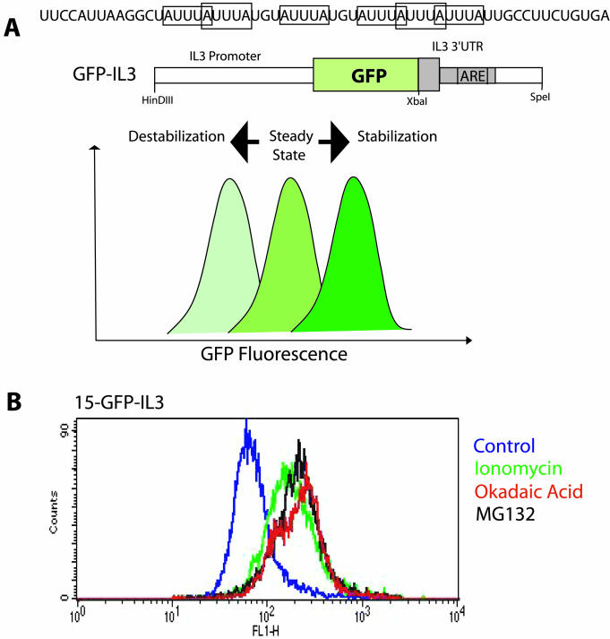
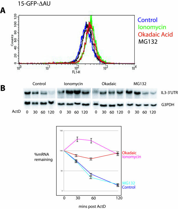
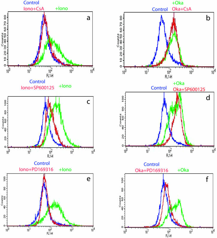
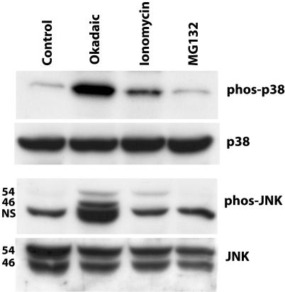
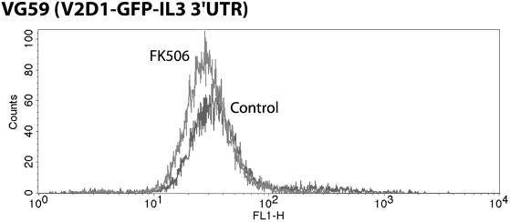
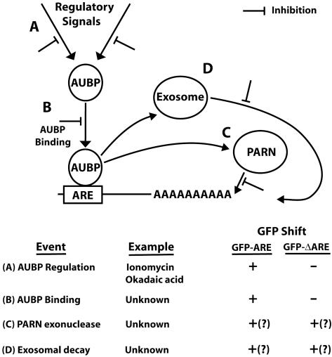
A reporter transcript containing the green fluorescent protein (GFP) gene upstream of the destabilizing 3'-untranslated region (3'-UTR) of the murine IL-3 gene was inserted in mouse PB-3c-15 mast cells. The GFP-IL-3 transcript was inherently unstable due to the presence of an adenosine-uridine (AU)-rich element (ARE) in the 3'-UTR and was subject to rapid decay giving a low baseline of GFP fluorescence. Transcript stabilization with ionomycin resulted in an increase of fluorescence that is quantitated by FACS analysis of responding cells. Using this system we have identified okadaic acid as a novel stabilizing compound, and investigated the upstream signaling pathways leading to stabilization. This reporter system has the advantage of speed and simplicity over standard methods currently in use and in addition to serving as a research tool it can be easily automated to increase throughput for drug discovery.


Figures

Figure 1
Figure 1
(A) The 59 nt long ARE region of the mouse IL-3 3′-UTR with AUUUA pentamers shown in boxes. A schematic representation of the GFP–IL3 reporter construct and the expected FACS profiles under conditions of mRNA stabilization and destabilization. (B) A representative FACS profile of 15-GFP–IL3 cells after treatment with ionomycin (1 μM), okadaic acid (400 nM) and MG132 (500 nM) for 4 h showing a substantial increase in GFP fluorescence.
Figure 2
Figure 2
(A) A representative FACS profile of the control non-ARE cell line, 15-GFP-ΔAU, showing no significant increase in GFP fluorescence after similar treatments with ionomycin, okadaic acid and MG132 as for the reporter cell line 15-GFP–IL3. (B) Actinomycin D chase mRNA decay assay of 15-GFP–IL3 cells over a 2 h time course. The blots were probed for the GFP–IL3 3′-UTR reporter to determine the rate of decay, then stripped and re-probed for the stable G3PD transcript for loading controls and to normalize the GFP–IL3 hybridization signals for quantification. Quantified results (±SE) for four independent experiments are shown on the graph. As shown, ionomycin and okadaic acid stabilize the reporter mRNA while MG132 is ineffective.
Figure 3
Figure 3
Antagonization of ionomycin-induced mRNA stabilization by cyclosporin A (0.5 μg/ml), PD169316 (10 μM) and SP600125 (10 μM). In contrast, okadaic acid-induced stabilization was uninhibited by cyclosporin A and SP600125 but was also sensitive to PD169316.
Figure 4
Figure 4
Western blots of 15-GFP–IL3 lysates treated with ionomycin, okadaic acid and MG132 for 30 min. Activation of the p38 MAPK pathway was determined with an antibody directed against the phosphorylated, active form of p38. JNK pathway activation was determined with a similar phospho-specific antibody that detects two phosphorylated isoforms of JNK (46 and 54 kDa), and a non-specific (NS) band. Blots were stripped and re-probed with antibodies against the unphosphorylated forms of the proteins as loading controls.
Figure 5
Figure 5
FACS profile of VG59 cells treated with FK506 (1 μg/ml) for 72 h.
Figure 6
Figure 6
Overview of the steps in mRNA decay. (A) Regulatory signals act on the AUBP to determine binding ability and hence stability/decay of the target transcript; (B) Binding of the AUBP to the ARE; (C) Deadenylation of the poly(A) tail by poly(A) ribonuclease (PARN); (D) 3′–5′ degradation of the deadenylated transcript by the cytoplasmic exosome. Decay can be potentially blocked by inhibition at any specific step. The table shows the expected results for different classes of inhibitors when screened using a GFP–ARE and the complementary ARE-deleted GFP–ΔARE cell lines. Question marks in parenthesis denotes where a particular interaction is hypothetical.

References

    1. Mitchell P. and Tollervey,D. (2000) mRNA stability in eukaryotes. Curr. Opin. Genet. Dev., 10, 193–198. - PubMed
    1. Bakheet T., Williams,B.R. and Khabar,K.S. (2003) ARED 2.0: an update of AU-rich element mRNA database. Nucleic Acids Res., 31, 421–3. - PMC - PubMed
    1. Guhayinogi J. and Brewer,G. (2001) Regulation of mRNA stability in mammalian cells. Gene, 265, 11–23. - PubMed
    1. Nair A.P.K., Hirsch,H.H. and Moroni,C. (1992) Mast cells sensitive to v-H-ras transformation are hyperinducible for IL-3 expression and have lost tumor-suppressor activity. Oncogene, 7, 1963–1972. - PubMed
    1. Stoecklin G., Ming,X.F., Looser,R. and Moroni,C. (2000) Somatic mRNA turnover mutants implicate tristetraprolin in the IL-3 mRNA degradation pathway. Mol. Cell. Biol., 20, 3753–3763. - PMC - PubMed

Publication types