. 2004 Jul 6;32(12):3531-6.
doi: 10.1093/nar/gkh676. Print 2004.

APE1 is the major 3'-phosphoglycolate activity in human cell extracts

Affiliations

APE1 is the major 3'-phosphoglycolate activity in human cell extracts

Jason L Parsons et al. Nucleic Acids Res. .

Abstract

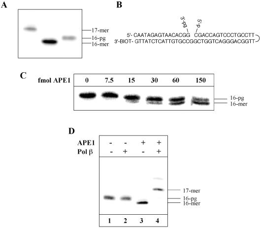
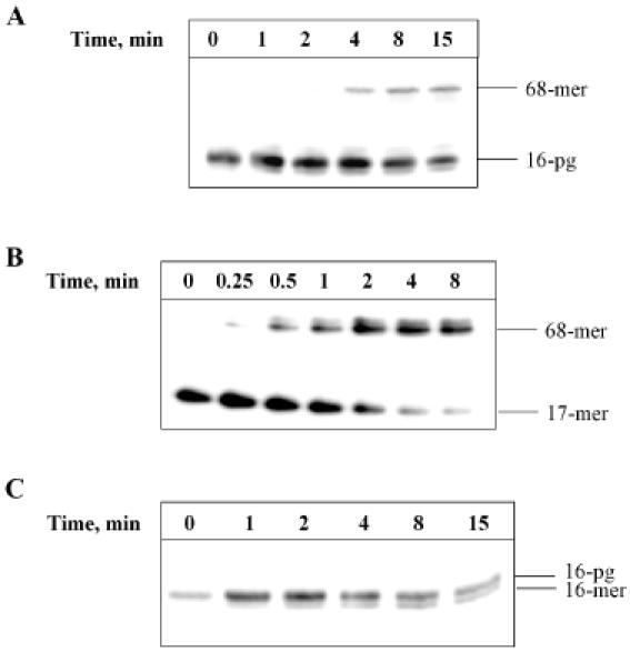
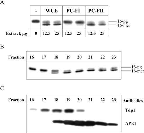
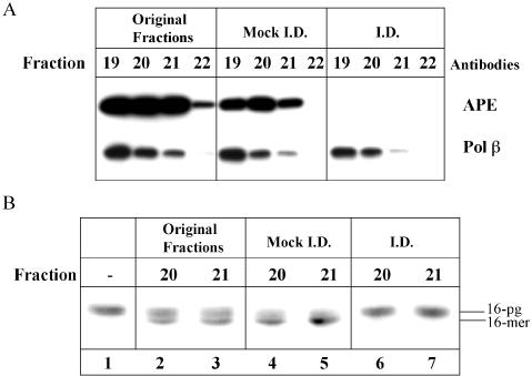
DNA strand breaks containing 3'-phosphoglycolate (3'-PG) ends are the major lesions induced by ionizing radiation. The repair of this lesion is not completely understood and several activities are thought to be involved in processing of 3'-PG ends. In this study we examined activities in human whole cell extracts (WCE) responsible for removal of 3'-PG. Using a radiolabelled oligonucleotide containing a single nucleotide gap with internal 5'-phosphate and 3'-PG ends, we demonstrate that the major 3'-PG activity in human WCE is Mg2+ dependent and that this activity co-purifies with AP endonuclease 1 (APE1) over phosphocellulose and gel filtration chromatography. Furthermore, immunodepletion of APE1 from active gel filtration fractions using APE1 specific antibodies reveals that the major activity against 3'-PG in human WCE is APE1.


Figures

Figure 1
Figure 1
Characterization of the 3′-PG-containing oligonucleotide substrate. An oligonucleotide (16mer) containing a 3′-PG end was 5′ end labelled and compared by 20% denaturing PAGE to the parent 16mer and a 1 nt addition product 17mer (A). The 5′ end labelled 3′-PG oligonucleotide was annealed to an oligonucleotide (51mer) containing a hairpin loop and a 5′-phosphate residue to generate a substrate for use in repair assays (B). This substrate was incubated with APE1 (0–150 fmol) for 10 min at 30°C prior to the addition of formamide loading dye. An aliquot was analysed by 20% denaturing PAGE and phosphorimaging (C). Primer extension from 3′-PG ends by human Pol β (50 fmol) was also investigated in the presence and absence of APE1 (150 fmol) (D).
Figure 2
Figure 2
Repair of a 3′-PG-containing duplex oligonucleotide by human WCE. The 5′ end labelled 3′-PG oligonucleotide was annealed to an oligonucleotide (51mer) containing a hairpin loop and a 5′-phosphate residue and bound to streptavidin beads prior to incubation with 100 μg WCE for 0–15 min at 30°C. Reactions were stopped by the addition of 100 mM EDTA, the beads washed and the DNA resuspended in formamide loading dye prior to analysis by 20% denaturing PAGE and phosphorimaging (A). Repair of a 5′-phosphate nick-containing oligonucleotide was observed by incubation of the substrate with 100 μg WCE for 0–8 min at 30°C (B). 3′-PG specific activity in WCE was observed by conducting reactions of the 3′-PG duplex oligonucleotide substrate with WCE in the absence of dNTPs (C).
Figure 3
Figure 3
Purification of 3′-PG activity from human WCE using phosphocellulose and gel filtration chromatography. WCE was loaded on a phosphocellulose column and fractions were step eluted using 0.15 M (PC-FI) and 1 M (PC-FII) KCl. Fractions were tested for 3′-PG activity using a 5′ end labelled 3′-PG oligonucleotide annealed to an oligonucleotide (51mer) containing a hairpin loop and a 5′-phosphate residue. The oligonucleotide substrate was bound to streptavidin beads prior to incubation with WCE or phosphocellulose fractions (12.5 and 25 μg) in the absence of dNTPs for 10 min at 30°C. The beads were washed and the DNA subsequently resuspended in formamide loading dye and analysed by 20% denaturing PAGE and phosphorimaging (A). Proteins from PC-FII were further separated by gel filtration on a Superdex 75 column and the fractions obtained were analysed for 3′-PG activity using the 3′-PG duplex oligonucleotide substrate in the absence of dNTPs by 20% denaturing PAGE and phosphorimaging (B). Aliquots of the fractions were also analysed by SDS–PAGE and western blotting using APE1 and Tdp1 antibodies (C).
Figure 4
Figure 4
Immunoprecipitation of APE1 from gel filtration fractions containing 3′-PG activity. Fractions from gel filtration chromatography containing 3′-PG activity (fractions 19–22) were mock-immunodepleted and immunodepleted of APE1 using APE1-specific antibodies. Samples were analysed by SDS–PAGE and western blotting using antibodies against APE1 and Pol β (A). The original fractions, mock-immunodepleted and immunodepleted fractions 20 and 21 were tested for 3′-PG activity using a 5′ end labelled 3′-PG oligonucleotide annealed to an oligonucleotide (51mer) containing a hairpin loop and a 5′-phosphate residue. Samples were incubated for 10 min at 30°C in the absence of dNTPs prior to the addition of formamide loading dye and analysis by 20% denaturing PAGE and phosphorimaging (B).

References

    1. Lindahl T. (1993) Instability and decay of the primary structure of DNA. Nature, 362, 709–715. - PubMed
    1. Dedon P.C. and Goldberg,I.H. (1992) Free-radical mechanisms involved in the formation of sequence-dependent bistranded DNA lesions by the antitumor antibiotics bleomycin, neocarzinostatin, and calicheamicin. Chem. Res. Toxicol., 5, 311–332. - PubMed
    1. Henle E.S. and Linn,S. (1997) Formation, prevention, and repair of DNA damage by iron/hydrogen peroxide. J. Biol. Chem., 272, 19095–19098. - PubMed
    1. Demple B. and DeMott,M.S. (2002) Dynamics and diversions in base excision DNA repair of oxidized abasic lesions. Oncogene, 21, 8926–8934. - PubMed
    1. Kane C.M. and Linn,S. (1981) Purification and characterization of an apurinic/apyrimidinic endonuclease from HeLa cells. J. Biol. Chem., 256, 3405–3414. - PubMed

MeSH terms