. 2004 Jul 20;101(29):10732-7.
doi: 10.1073/pnas.0404117101. Epub 2004 Jul 9.

Vasorin, a transforming growth factor beta-binding protein expressed in vascular smooth muscle cells, modulates the arterial response to injury in vivo

Affiliations

Vasorin, a transforming growth factor beta-binding protein expressed in vascular smooth muscle cells, modulates the arterial response to injury in vivo

Yuichi Ikeda et al. Proc Natl Acad Sci U S A. .

Abstract

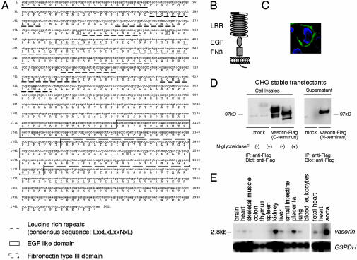
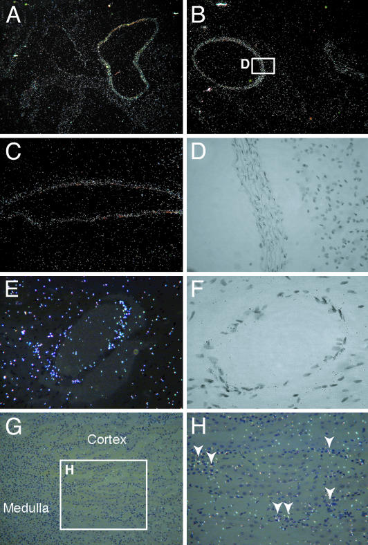
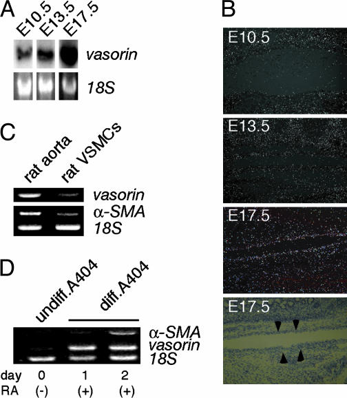
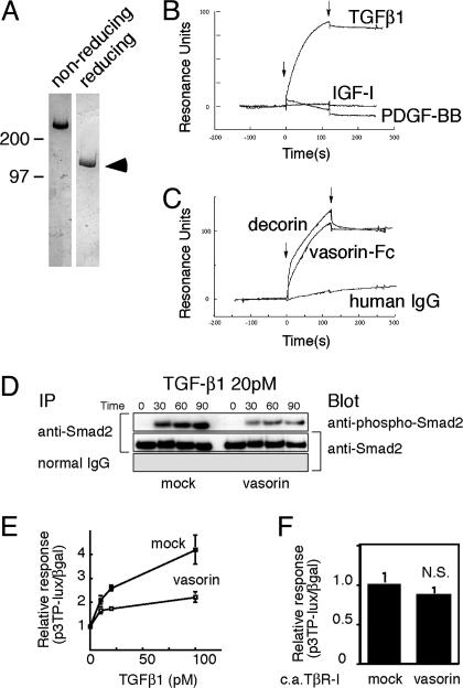
Growth factors, cell-surface receptors, adhesion molecules, and extracellular matrix proteins play critical roles in vascular pathophysiology by affecting growth, migration, differentiation, and survival of vascular cells. In a search for secreted and cell-surface molecules expressed in the cardiovascular system, by using a retrovirus-mediated signal sequence trap method, we isolated a cell-surface protein named vasorin. Vasorin is a typical type I membrane protein, containing tandem arrays of a characteristic leucine-rich repeat motif, an epidermal growth factor-like motif, and a fibronectin type III-like motif at the extracellular domain. Expression analyses demonstrated that vasorin is predominantly expressed in vascular smooth muscle cells, and that its expression is developmentally regulated. To clarify biological functions of vasorin, we searched for its binding partners and found that vasorin directly binds to transforming growth factor (TGF)-beta and attenuates TGF-beta signaling in vitro. Vasorin expression was down-regulated during vessel repair after arterial injury, and reversal of vasorin down-regulation, by using adenovirus-mediated in vivo gene transfer, significantly diminished injury-induced vascular lesion formation, at least in part, by inhibiting TGF-beta signaling in vivo. These results suggest that down-regulation of vasorin expression contributes to neointimal formation after vascular injury and that vasorin modulates cellular responses to pathological stimuli in the vessel wall. Thus, vasorin is a potential therapeutic target for vascular fibroproliferative disorders.


Figures

Fig. 1.
Fig. 1.
Vasorin, an identified cell-surface protein. (A) Deduced amino acid sequence of human vasorin. The putative signal peptide (underlined), the LRRs (dotted underlines), the EGF motif (boxed), the fibronectin type III motif (dotted boxes), the transmembrane sequence (underlined), and five putative N-glycosylation sites (boxed) are indicated. (B) Structural model of vasorin. (C) Immunofluorescence analysis of subcellular localization of vasorin. Vasorin was expressed on the cell surface. (D) Cell lysates and supernatants of CHO cells stably expressing vasorin-Flag were subjected to immunoprecipitation and Western blot analysis by using an anti-Flag antibody. An ≈110-kDa protein for membrane-bound vasorin with C-terminal tag and a ≈100-kDa protein for soluble vasorin with N-terminal tag were detected under reducing conditions. N-glycosidase F treatment revealed that vasorin is N-glycosylated. (E) Northern blot analysis of adult human tissues. A single intense 2.8-kb band was detected and the strongest expression was observed in the aorta.
Fig. 2.
Fig. 2.
In situ hybridization analysis of vasorin. Sections of adult mouse aorta at different levels (A–D), the coronary artery (E and F), and the kidney (G and H) are shown. Vasorin is expressed in VSMCs of different origins. White spots represent hybridization signals. (A) The proximal ascending aorta. (B) The descending thoracic aorta. (C) The abdominal aorta. (D) Partial magnification of bright-field image of B. Black spots within the elastic fibers represent hybridization signals. (E) The coronary artery. (F) A bright-field image of the coronary artery. Black spots represent hybridization signals. (G) The kidney. (H) Partial magnification of G. Vasorin is expressed in interstitial cells.
Fig. 3.
Fig. 3.
Developmental regulation of vasorin. (A) Northern blot analysis of staged mouse embryos (E10.5, E13.5, and E17.5). (B) Expression pattern of vasorin during aortic development examined by in situ hybridization analyses. The fourth image is the corresponding bright-field image of the third representation. Arrowheads indicate the aorta in the mouse embryo (E17.5). (C) Semiquantitative RT-PCR comparing the expression of vasorin in the adult rat aorta with that in cultured rat aortic VSMCs. Rat α-smooth muscle actin (α-SMA) and 18S rRNA were used as a positive and an internal control, respectively. (D) Semiquantitative RT-PCR showing the induction of the vasorin gene in RA-treated A404 cells. Rat α-SMA and 18S rRNA were used as a positive and an internal control, respectively.
Fig. 4.
Fig. 4.
Vasorin directly binds to TGF-β and modulates TGF-β signaling in vitro. (A) Purified recombinant vasorin-Fc fusion protein was free of protein contamination, as estimated by SDS/PAGE, followed by Coomassie blue staining. (B) Sensorgrams obtained from injection of vasorin-Fc on immobilized TGF-β1, PDGF-BB, and insulin-like growth factor I (IGF-I) are shown. (C) Sensorgrams obtained from injection of vasorin-Fc, decorin, and human IgG on immobilized TGF-β1 are shown. Arrowheads indicate initiation and termination of injections. (D) TGF-β-induced Smad2 phosphorylation was significantly inhibited in vasorin-expressing cells. Stable transfectants were treated with TGF-β1 (20 pM), and then immunoprecipitated with an anti-Smad2/3 antibody, followed by blotting with an anti-phospho-Smad2 antibody. (E) A reporter assay was performed by using the TGF-β-responsive reporter p3TP-lux. Stable transfectants were stimulated with TGF-β1 at various concentrations, and vasorin inhibited TGF-β-induced reporter gene activation. (F) Vasorin inhibited TGF-β signaling at the extracellular and/or cell-surface level. The p3TP-lux reporter and the constitutively active TβR-I were cotransfected into stable transfectants. Transfection of the constitutively active TβR-I activated the p3TP-lux reporter, but vasorin did not significantly inhibit this activation. N.S., not significant.
Fig. 5.
Fig. 5.
Vasorin expression was down-regulated during vessel repair after arterial injury, and reversal of vasorin down-regulation significantly reduced neointimal formation, at least in part, by attenuating TGF-β signaling in vivo. (A) Rat carotid arteries were harvested at 3 days after injury to examine the expression levels of vasorin by semiquantitative RT-PCR analysis. Down-regulation of vasorin expression was induced by mechanical vascular injury, and Ad-vasorin treatment partially reversed this down-regulation. In contrast to vasorin, the expression of TGF-β1, TNF-α, and IL-6 was up-regulated by vascular injury and was not altered by vasorin administration. GAPDH was used as an internal control. (B) Vessels treated with Ad-vasorin were harvested 3 days afterward and were subjected to immunostaining to confirm protein expression by using the anti-Flag antibody. (C) Effects of Ad-vasorin on neointimal formation in rat carotid arteries at 14 days after injury (n = 5 arteries for each group). Representative hematoxylin/eosin-stained cross sections (Top and Middle) and Sirius red-stained cross sections (Bottom) of balloon-injured arteries treated with PBS (Left), Ad-β-galactosidase (Center), and Ad-vasorin (Right) are shown. (Middle) Partial magnifications of the respective Top images. Ad-vasorin administration significantly reduced the intima/media area ratio of injured arteries and collagen content in the lesions (P < 0.01), as compared with Ad-β-galactosidase administration. NI, neointima; M, media; HE, hematoxylin/eosin. *, P < 0.05; **, P < 0.01. (D) The inhibitory effects of vasorin administration on TGF-β signaling in vivo. Arteries were harvested 3 days after injury and were subjected to Western blot analysis by using the anti-phospho-Smad2 antibody. Representative data are shown. Smad2 phosphorylation was significantly reduced in all Ad-vasorin-treated arteries (P < 0.05). The blots were stripped and reprobed with the anti-α-tubulin antibody to ensure equal loading of proteins. The relative intensities of phospho-Smad2 bands were measured by densitometric scanning from three independent experiments. *, P < 0.05.

References

    1. Tashiro, K., Tada, H., Heilker, R., Shirozu, M., Nakano, T. & Honjo, T. (1993) Science 261, 600-603. - PubMed
    1. Klein, R. D., Gu, Q., Goddard, A. & Rosenthal, A. (1996) Proc. Natl. Acad. Sci. USA 93, 7108-7113. - PMC - PubMed
    1. Kojima, T. & Kitamura, T. (1999) Nat. Biotechnol. 17, 487-490. - PubMed
    1. Nosaka, T., Kawashima, T., Misawa, K., Ikuta, K., Mui, A. L. & Kitamura, T. (1999) EMBO J. 18, 4754-4765. - PMC - PubMed
    1. Chamley-Campbell, J., Campbell, G. R. & Ross, R. (1979) Physiol. Rev. 59, 1-61. - PubMed

Publication types

MeSH terms

Substances

Associated data