. 2004 Jul 20;101(29):10673-8.
doi: 10.1073/pnas.0403139101. Epub 2004 Jul 9.

NFAT5/TonEBP mutant mice define osmotic stress as a critical feature of the lymphoid microenvironment

Affiliations

NFAT5/TonEBP mutant mice define osmotic stress as a critical feature of the lymphoid microenvironment

William Y Go et al. Proc Natl Acad Sci U S A. .

Abstract

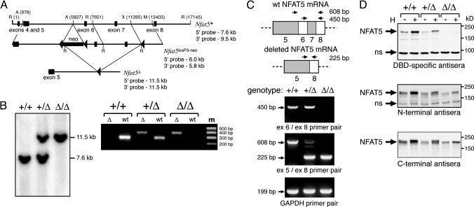
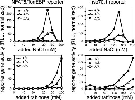
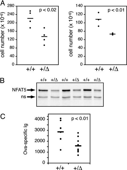
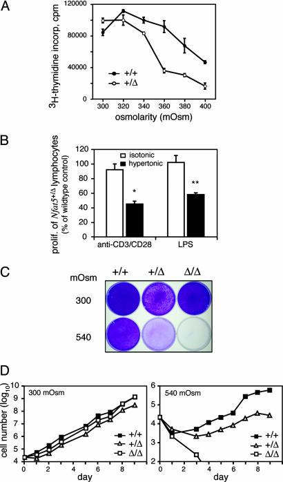
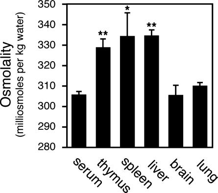
Osmotic stress responses are critical not only to the survival of unicellular organisms but also to the normal function of the mammalian kidney. However, the extent to which cells outside the kidney rely on osmotic stress responses in vivo remains unknown. Nuclear factor of activated T cells 5 (NFAT5)/tonicity enhancer binding protein (TonEBP), the only known osmosensitive mammalian transcription factor, is expressed most abundantly in the thymus and is induced upon lymphocyte activation. Here we report that NFAT5/TonEBP is not only essential for normal cell proliferation under hyperosmotic conditions but also necessary for optimal adaptive immunity. Targeted deletion of exons 6 and 7 of the Nfat5 gene, which encode a critical region of the DNA-binding domain, gave rise to a complete loss of function in the homozygous state and a partial loss of function in the heterozygous state. Complete loss of function resulted in late gestational lethality. Furthermore, hypertonicity-induced NFAT5/TonEBP transcriptional activity and hsp70.1 promoter function were completely eliminated, and cell proliferation under hyperosmotic culture conditions was markedly impaired. Partial loss of NFAT5/TonEBP function resulted in lymphoid hypocellularity and impaired antigen-specific antibody responses in viable heterozygous animals. In addition, lymphocyte proliferation ex vivo was reduced under hypertonic, but not isotonic, culture conditions. Direct measurement of tissue osmolality further revealed lymphoid tissues to be hyperosmolar. These results indicate that lymphocyte-mediated immunity is contingent on adaptation to physiologic osmotic stress, thus providing insight into the lymphoid microenvironment and the importance of the NFAT5/TonEBP osmotic stress response pathway in vivo.


Figures

Fig. 1.
Fig. 1.
The targeted deletion of exons 6 and 7 of the Nfat5 gene results in expression of a mutant NFAT5 protein. (A) Gene-targeting strategy for deletion of exons 6 and 7 of the Nfat5 gene by homologous recombination in ES cells (see Materials and Methods). A, AvrII; R, EcoRI; M, MscI; X, XhoI. (B) Targeted deletion within the Nfat5 gene was verified by PCR genotyping with two sets of primers that span the site of insertion of the downstream loxP site in both the wild-type and knockout alleles (Left), and by Southern blot analysis with a 5′ probe located outside of the targeted genomic sequence (Right). (C) RNA isolated from SV40 T antigen-immortalized MEF cell lines was subjected to RT-PCR analysis with primers positioned as indicated relative to that portion of the NFAT5 mRNA encoded by exons 5–8. (D) Whole-cell extracts from MEF cell lines cultured under either standard or hypertonic (H; complete medium plus additional 120 mM NaCl) culture conditions for 16 h were subjected to immunoblot analysis with the indicated antisera. The indicated nonspecific (ns) bands function as internal controls for equal protein loading and transfer.
Fig. 2.
Fig. 2.
The Nfat5Δ allele confers either partial or complete loss of NFAT5 function. Immortalized MEF cell lines derived from Nfat5+/+, Nfat5+/Δ, and Nfat5Δ/Δ embryos were transfected with the indicated reporter gene. Approximately 24 h after transfection, the cells were cultured in complete medium with either NaCl or raffinose added to the indicated concentration. Cell extracts were prepared 16 h later for assay of reporter activity. The results represent luciferase reporter activity (relative light units, RLU) normalized to correct for variation in transfection efficiency and are representative of three independent experiments.
Fig. 3.
Fig. 3.
Partial loss of NFAT5 function results in impaired adaptive immunity. (A) The cellularity of the thymus (Left) and spleen (Right) from 5-week-old male Nfat5+/+ and Nfat5+/Δ littermates was determined by performing manual cell counts. The viable cells were distinguished by using trypan blue dye exclusion. (B) Whole-cell extracts of thymocytes from Nfat5+/Δ mice and Nfat5+/+ littermate controls were prepared and probed for NFAT5 expression by Western blot analysis with the C-terminal NFAT5 antisera. The indicated nonspecific (ns) band provides an internal control for equal protein loading and transfer. (C) Relative antigen-specific Ig levels were measured by ELISA of serum from Nfat5+/Δ and Nfat5+/+ mice collected 21 days after immunization with ovalbumin.
Fig. 4.
Fig. 4.
Normal cell proliferation under conditions of hyperosmotic stress requires NFAT5. (A) The splenocytes from Nfat5+/Δ and Nfat5+/+ littermates (5–8 weeks old) were cultured in standard complete medium (≈290 mOsm) or complete medium supplemented with raffinose to increase the osmolality of the culture conditions as indicated. The cells were stimulated with anti-CD3 plus anti-CD28 to induce T cell proliferation. (B) The splenocytes from Nfat5+/Δ mice and Nfat5+/+ littermate controls were cultured in standard complete medium (≈290 mOsm) or subjected to hypertonic stress (≈370 mOsm) through the addition of 80 mM raffinose. The cells were stimulated in the previously described method to induce T cell proliferation (n = 7 Nfat5+/+ and Nfat5+/Δ littermate pairs) or with LPS to induce B cell proliferation (n = 3 littermate pairs). Statistically significant differences are indicated (*, P < 0.01; **, P < 0.0001). (C) Immortalized MEF cell lines were cultured in complete medium adjusted to 300 mOsm in the absence and presence of additional NaCl (120 mM). The cells were seeded into the indicated medium and allowed to grow for 6 days, during which time the cells grown in standard medium were split and replated once. The plates were fixed and stained with crystal violet dye. (D) The MEF cell lines were seeded in 24-well tissue culture dishes and allowed to grow until ≈80% confluence, at which time the cultures were continued by replating. Manual cell counts were performed on the indicated days by using trypan blue exclusion to identify viable cells. The culture medium was replaced every 3 days. The data shown represent the mean of cell counts from triplicate wells. Standard errors, which were <5% of the mean, are not shown. These results are representative of at least four independent measurements of cell growth.
Fig. 5.
Fig. 5.
Hyperosmolality of lymphoid tissues relative to blood. Tissue osmolality was determined by vapor pressure osmometry (see Materials and Methods). Statistically significant differences between blood and tissue osmolality are indicated (*, P < 0.02; **, P < 0.001).

References

    1. Wood, J. M., Bremer, E., Csonka, L. N., Kraemer, R., Poolman, B., van der Heide, T. & Smith, L. T. (2001) Comp. Biochem. Physiol. A Physiol. 130, 437–460. - PubMed
    1. Hohmann, S. (2002) Microbiol. Mol. Biol. Rev. 66, 300–372. - PMC - PubMed
    1. Yancey, P. H., Clark, M. E., Hand, S. C., Bowlus, R. D. & Somero, G. N. (1982) Science 217, 1214–1222. - PubMed
    1. Burg, M. B. (1995) Am. J. Physiol. 268, F983–F996. - PubMed
    1. Miyakawa, H., Woo, S. K., Dahl, S. C., Handler, J. S. & Kwon, H. M. (1999) Proc. Natl. Acad. Sci. USA 96, 2538–2542. - PMC - PubMed

Publication types

MeSH terms

Substances