. 2004 Aug;5(8):812-7.
doi: 10.1038/sj.embor.7400190. Epub 2004 Jul 9.

UVA inactivates protein tyrosine phosphatases by calpain-mediated degradation

Affiliations

UVA inactivates protein tyrosine phosphatases by calpain-mediated degradation

Pawan Gulati et al. EMBO Rep. 2004 Aug.

Erratum in

  • EMBO Rep. 2005 Nov;6(11):1101

Abstract

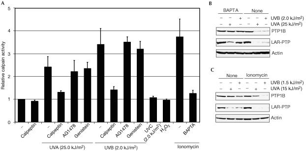
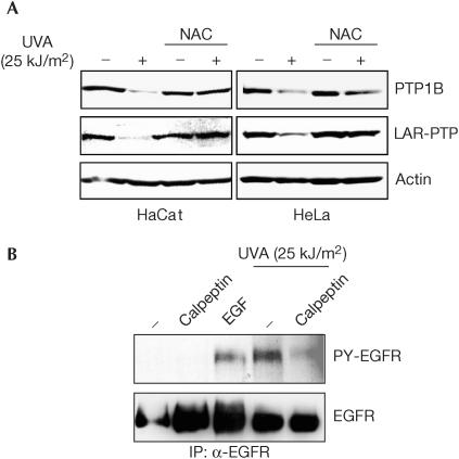
UV irradiation causes inflammatory and proliferative cellular responses. We have proposed previously that these effects are, to a large extent, caused by the ligand-independent activation of several receptor tyrosine kinases due to the inactivation of their negative control elements, the protein tyrosine phosphatases (PTPs). We examined the mechanism of this inactivation and found that, in addition to reversible oxidation of PTPs, UV triggers a novel mechanism: induced degradation of PTPs by calpain, which requires both calpain activation and substrate PTP oxidative modification. This as yet unrecognized effect of UV is irreversible, occurs predominantly with UVA and UVB, the range of wavelengths in sunlight that reach the skin surface, and at physiologically relevant doses.


Figures

Figure 1
Figure 1
Loss of protein tyrosine phosphatase activity and protein from A431 cells by UV irradiation. (A) Hypersensitivity of PTP1B to UV irradiation. Confluent A431 cells were irradiated with different doses of UVA. PTP1B was immunoprecipitated from lysates, and its activity was measured using the pNPP assay. Alkaline phosphatase (AP) and alcohol dehydrogenase (ADH) activities were assayed as described in Methods, and the activity of caspase 3 was assayed using a commercial kit (Calbiochem). (B) Reversible inactivation of PTP1B. PTP1B immunoprecipitates obtained as in (A) were split, either mock-treated or treated with 50 mM NAC at 25°C for 10 min under agitation, and activity was determined by the pNPP assay. (C) The data shown in (B) were normalized for the protein amounts of PTP1B in the immunoprecipitates as detected by immunoblotting. (D) Loss of PTP protein on UV irradiation. Within 1–2 min after irradiation, cells were lysed and subjected to western blotting using antibodies directed against PTP1B, SHP1, LAR-PTP and actin. (E) No loss of PP-2A, pro-caspase 8, actin or Hsp90 occurred on UV irradiation. Immunoblots are as in (D).
Figure 2
Figure 2
Calpain-dependent protein tyrosine phosphatase degradation. (A) Active fragments of PTP1B were detected by in-gel assay (Burridge & Nelson, 1995; Markova et al, 2003). (B,C) The calpain inhibitor calpeptin prevents UV-induced degradation of PTPs. PTP1B was immunoprecipitated before western blotting, and LAR-PTP was blotted directly from the lysate. Before UV irradiation, A431 cells were mock-treated or were treated with calpeptin (1 μM) at 37°C for 4 h. Cell lysates of all samples were generated in the presence of calpeptin (5 μM).
Figure 3
Figure 3
UV induces calcium-dependent calpain activation and protein tyrosine phosphatase degradation. (A) Relative calpain activity after various types of treatment. A431 cells were pretreated with calpeptin as in Fig 2B,C. AG1478 (100 nM), genistein (100 μM) or H2O2 (5 mM) was present for 15 min, ionomycin (2 μM) for 2 min or BAPTA (2 μM) for 30 min before UV irradiation. Calpain activity of lysates was determined using a commercially available kit (Calbiochem). The activity of untreated cells, set at 1, represents cleavage of 53 pmol/min of substrate at 37°C. (B,C) Modulation of intracellular calcium levels affects UV-induced PTP degradation. (B) Cell-permeable calcium chelator blocks UV-induced PTP degradation. A431 cells were cultivated in calcium-free medium for 30 min, and then treated with BAPTA (2 μM) for 30 min before irradiation. Immunoblots of lysates are shown. (C) Ionomycin enhances UV-induced PTP degradation. Ionomycin treatment is as in (A); immunoblots of lysates are shown.
Figure 4
Figure 4
UV- and calpain-dependent protein tyrosine phosphatase degradation requires previous PTP oxidation. (A) Antioxidant treatment attenuates UV-induced degradation. A431 cells were treated with NAC (30 mM) for 2 min before irradiation, and the lysates were immunoblotted. (B) Reversible oxidation converts PTP1B to a calpain substrate in vitro. Left panel: Cells were treated with H2O2 (5 mM) at 37°C for 15 min and PTP1B was immunoprecipitated. The precipitates were divided into two parts, one of which was digested with 0.4 U of calpain at 37°C for 30 min. Right panel: PTP1B was immunoprecipitated from an untreated cell lysate, and either reversibly or irreversibly oxidized by treatment with 50 μM of H2O2 for 4 or 16 h, respectively (Salmeen et al, 2003; supplementary Fig 3 online) and exposed to calpain as in the left panel. (C) Reversible oxidation of PTP1B increases the susceptibility to calpain but not to other proteinases. PTP1B was immunoprecipitated and reversibly oxidized (as in Fig 3B) or mock-treated. The immunoprecipitates were then left untreated or were treated with 0.4 U of the indicated proteinases at 37°C for different periods. PTP1B protein digestion was measured by quantification of the PTP1B band in immunoblots. The ratio of the amount of reversibly oxidized versus non-oxidized PTP1B remaining after digestion is depicted. (D) Combined treatment with H2O2 (as in (B)) and ionomycin (as in Fig 3A) leads to PTP degradation. Immunoblots of lysates are shown.
Figure 5
Figure 5
Physiological significance of UVA induced PTP degradation. (A) Generality of UVA-induced PTP degradation. HaCat and HeLa cells were left untreated or were treated with NAC as in Fig 4A, before mock treatment or UVA irradiation, and the lysates were immunoblotted. (B) UVA-induced activation of epidermal growth factor receptor (EGFR) requires calpain activity. A431 cells were treated with calpeptin before irradiation as in Fig 2 or stimulated with EGF (50 ng/ml) at 37°C for 5 min, where indicated. EGFR was immunoprecipitated from lysates and its tyrosine phosphorylation was determined by immunoblotting.

References

    1. Andersen JN, Mortensen OH, Peters GH, Drake PG, Iversen LF, Olsen OH, Jansen PG, Andersen HS, Tonks NK, Moller NP (2001) Structural and evolutionary relationships among protein tyrosine phosphatase domains. Mol Cell Biol 21: 7117–7136 - PMC - PubMed
    1. Aragane Y, Kulms D, Metze D, Wilkes G, Poppelmann B, Luger TA, Schwarz T (1998) Ultraviolet light induces apoptosis via direct activation of CD95 (Fas/APO-1) independently of its ligand CD95L. J Cell Biol 140: 171–182 - PMC - PubMed
    1. Barrett WC, DeGnore JP, Konig S, Fales HM, Keng YF, Zhang ZY, Yim MB, Chock PB (1999) Regulation of PTP1B via glutathionylation of the active site cysteine 215. Biochemistry 38: 6699–6705 - PubMed
    1. Burridge K, Nelson A (1995) An in-gel assay for protein tyrosine phosphatase activity: detection of widespread distribution in cells and tissues. Anal Biochem 232: 56–64 - PubMed
    1. Coffer PJ, Burgering BM, Peppelenbosch MP, Bos JL, Kruijer W (1995) UV activation of receptor tyrosine kinase activity. Oncogene 11: 561–569 - PubMed

Publication types