. 2004 Jul 12:4:27.
doi: 10.1186/1471-2180-4-27.

Yersinia enterocolitica type III secretion: evidence for the ability to transport proteins that are folded prior to secretion

Affiliations

Yersinia enterocolitica type III secretion: evidence for the ability to transport proteins that are folded prior to secretion

Gottfried Wilharm et al. BMC Microbiol. .

Abstract

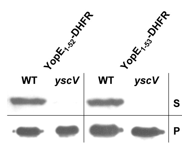
Background: Pathogenic Yersinia species (Y. enterocolitica, Y. pestis, Y. pseudotuberculosis) share a type three secretion system (TTSS) which allows translocation of effector proteins (called Yops) into host cells. It is believed that proteins are delivered through a hollow needle with an inner diameter of 2-3 nm. Thus transport seems to require substrates which are essentially unfolded. Recent work from different groups suggests that the Yersinia TTSS cannot accommodate substrates which are folded prior to secretion. It was suggested that folding is prevented either by co-translational secretion or by the assistance of specific Yop chaperones (called Sycs).

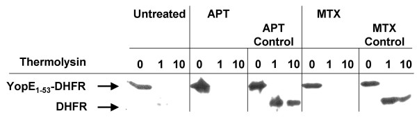
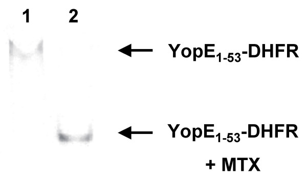
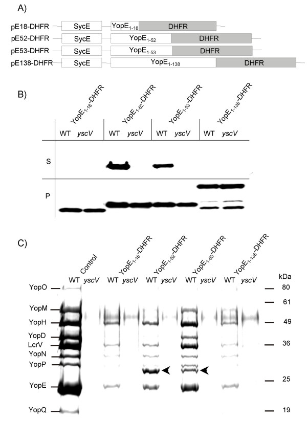
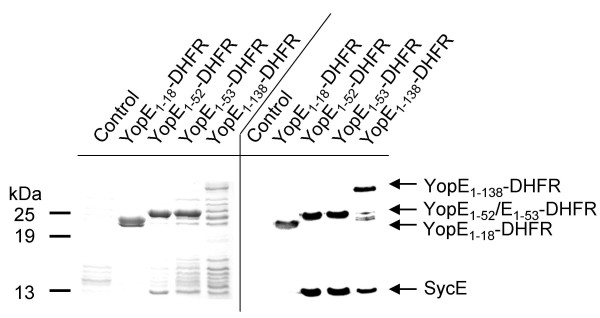
Results: In this study we have fused YopE secretion signals of various length to the mouse dihydrofolate reductase (DHFR) in order to analyse the DHFR folding state prior to secretion. We could demonstrate that secretion-deficient as well as secretion-competent YopE-DHFR fusions complexed to SycE can be efficiently purified from Yersinia cytosol by affinity chromatography using methotrexate-agarose. This implies the folding of the DHFR fusion moiety despite SycE binding and contradicts the previously presented model of folding inhibition by chaperone binding. Secretion-deficient YopE-DHFR fusions caused severe jamming of the TTSS. This observation contradicts the co-translational secretion model.

Conclusions: We present evidence that the Yersinia TTSS is familiar with the processing of transport substrates which are folded prior to secretion. We therefore predict that an unfoldase is involved in type III secretion.


Figures

Figure 1
Figure 1
Protease protection assay on cytosolic YopE1–53-DHFR prepared from spheroplasts. Spheroplasts were prepared from yersiniae induced for Yop expression and treated with 1 mM methotrexate (MTX), 1 mM aminopterin (APT) or untreated, respectively. Spheroplasts were washed three times and subsequently lysed by osmotic shock, then soluble supernatants after centrifugation (cytosolic fractions) were treated with thermolysin (0, 1, 10 μg/ml). As "control", cytosol from untreated yersiniae was incubated with 1 mM MTX and 1 mM APT, respectively, prior to thermolysin exposition. After incubation at 20°C for 15 minutes samples were loaded on an SDS gel that was electroblotted. Immunostaining was performed with anti-DHFR monoclonal antibodies.
Figure 2
Figure 2
Native gel electrophoresis reveals binding of methotrexate (MTX) to YopE1–53-DHFR. Cytosolic fractions from yersiniae expressing YopE1–53-DHFR were prepared as described in legend to Fig. 1. Cytosolic fraction was loaded directly on a HEPES-buffered native gel (lane 1) and after further incubation with 1 mM MTX (lane 2). After electrophoresis gel was electroblotted and DHFR-fusions were detected using anti-DHFR antibodies.
Figure 3
Figure 3
Expression and secretion of YopE-DHFR fusions. (A) Scheme depicts plasmid constructs pE18-DHFR, pE52-DHFR, pE53-DHFR and pE138-DHFR, allowing expression of YopE N-termini (the first 18, 52, 53 or 138 aa) fused to DHFR and concomitant expression of YopE-chaperone SycE. (B) Yersiniae (Y. enterocolitica strain WA-314, abbrev. WT, and its secretion deficient yscV mutant (also termed lcrD)) expressing YopE-DHFR fusions were cultured for 2 h at 37°C in BHI medium. Then Yop secretion was induced by Ca2+-depletion with EGTA. After 1.5 h of continued incubation bacteria were pelleted and whole cell lysates (P) and TCA-precipitated supernatants (S) were subjected to SDS-PAGE; ten times more supernatant than cell pellet was loaded. Subsequently, immunoblotting was performed with monoclonal anti-DHFR antibodies. (C) Coomassie-stained SDS-PAGE of supernatants from a secretion experiment as described above (Fig. 3B). The first two lanes show Yop secretion of the parental hosts. Arrows indicate secreted YopE1–52-DHFR and YopE1–53-DHFR.
Figure 4
Figure 4
Affinity purification of YopE-DHFR fusions using MTX-agarose. Yersiniae expressing DHFR fusions as indicated were lysed by French press treatment and soluble supernatants after centrifugation were incubated with MTX-agarose beads on ice for 30 min. Beads were washed with PBS five times, subsequently resuspended in SDS loading buffer and subjected to SDS-PAGE. Left panel represents a Coomassie stained gel, right panel a Western blot analysed with monoclonal anti-DHFR antiserum and polyclonal anti-SycE antiserum simultaneously. As control served Y. enterocolitica WA-314 harbouring plasmid pACYC184.
Figure 5
Figure 5
Secretion of YopE1–52-DHFR and YopE1–53-DHFR after inhibition of protein biosynthesis. Yersiniae (Y. enterocolitica strain WA-314, abbrev. WT, and the secretion deficient yscV mutant) expressing YopE1–52-DHFR and YopE1–53-DHFR, respectively, were cultured for 2 h at 37°C in BHI supplemented with CaCl2. Then 50 μg/ml of tetracycline was added to inhibit protein synthesis and cultures were incubated for another 20 min. Then bacteria were pelleted and resuspended in BHI depleted of Ca2+ to induce Yop secretion in the presence of tetracycline. After 20 min of continued incubation bacteria were pelleted and whole cell lysates (P) and TCA-precipitated supernatants (S) were subjected to SDS-PAGE; ten times more supernatant than cell pellet was loaded. Subsequently, immunoblotting was performed with monoclonal anti-DHFR antibodies.

Similar articles

Cited by

References

    1. Rosqvist R, Magnusson KE, Wolf-Watz H. Target cell contact triggers expression and polarized transfer of Yersinia YopE cytotoxin into mammalian cells. EMBO J. 1994;13:964–972. - PMC - PubMed
    1. Cornelis GR. The Yersinia Ysc-Yop virulence apparatus. Int J Med Microbiol. 2002;291:455–462. - PubMed
    1. Cornelis GR, Boland A, Boyd AP, Geuijen C, Iriarte M, Neyt C, Sory MP, Stainier I. The virulence plasmid of Yersinia, an antihost genome. Microbiol Mol Biol Rev. 1998;62:1315–1352. - PMC - PubMed
    1. Hoiczyk E, Blobel G. Polymerization of a single protein of the pathogen Yersinia enterocolitica into needles punctures eukaryotic cells. Proc Natl Acad Sci USA. 2001;98:4669–4674. doi: 10.1073/pnas.071065798. - DOI - PMC - PubMed
    1. Anderson DM, Schneewind O. A mRNA signal for the type III secretion of Yop proteins by Yersinia enterocolitica. Science. 1997;278:1140–1143. doi: 10.1126/science.278.5340.1140. - DOI - PubMed

Publication types

MeSH terms

LinkOut - more resources