. 2004 Jul 12;32(12):e100.
doi: 10.1093/nar/gnh098.

Measurement of DNA mismatch repair activity in live cells

Affiliations

Measurement of DNA mismatch repair activity in live cells

Xiufen Lei et al. Nucleic Acids Res. .

Abstract

Loss of DNA mismatch repair (MMR) function leads to the development and progression of certain cancers. Currently, assays for DNA MMR activity involve the use of cell extracts and are technically challenging and costly. Here, we report a rapid, less labor-intensive method that can quantitatively measure MMR activity in live cells. A G-G or T-G mismatch was introduced into the ATG start codon of the enhanced green fluorescent protein (EGFP) gene. Repair of the G-G or T-G mismatch to G-C or T-A, respectively, in the heteroduplex plasmid generates a functional EGFP gene expression. The heteroduplex plasmid and a similarly constructed homoduplex plasmid were transfected in parallel into the same cell line and the number of green cells counted by flow cytometry. Relative EGFP expression was calculated as the total fluorescence intensity of cells transfected with the heteroduplex construct divided by that of cells transfected with the homoduplex construct. We have tested several cell lines from both MMR-deficient and MMR-proficient groups using this method, including a colon carcinoma cell line HCT116 with defective hMLH1 gene and a derivative complemented by transient transfection with hMLH1 cDNA. Results show that MMR-proficient cells have significantly higher EGFP expression than MMR-deficient cells, and that transient expression of hMLH1 alone can elevate MMR activity in HCT116 cells. This method is potentially useful in comparing and monitoring MMR activity in live cells under various growth conditions.


Figures

Figure 1
Figure 1
Flow chart of protocol for the quantitation of DNA MMR in vivo. MMR efficiency as measured with relative EGFP expression was calculated using the equation of (M · IMN · IN)/(C · ICN · IN), where M, N or C is the percentage of green cells for heteroduplex, negative control or homoduplex transfection, respectively, and IM, IN or IC is the mean fluorescence intensity of green cells for M, N or C, respectively. PSAD stands for plasmid-safe ATP-dependent DNase.
Figure 2
Figure 2
Restriction analysis of ligation products. (A) Digestion of un-nicked homoduplex and heteroduplex plasmids with NcoI. The ligation products was digested with NcoI endonuclease and then electrophoresed in 1% agarose gel. The homoduplex plasmid was digested into three fragments of 2.3, 1.9 and 317 bp. The ethidium bromide staining of the 317 bp fragment was too weak to be photographed. A faint band at 2.6 kb is likely the incompletely digested product that generates 2.3 and 317 bp fragments. The heteroduplex plasmid was digested into two fragments of 2.6 and 1.9 kb. (B) Digestion of nicked homoduplex and heteroduplex plasmids with NcoI. Homoduplex was digested into three fragments of 3.3, 1.0 and 317 bp, whereas the heteroduplex was digested into two fragments of 3.3 and 1.3 kb.
Figure 3
Figure 3
Heteroduplex EGFP plasmid is effectively repaired in HeLa cell, but not in HCT116 cell. (A) Fluorescent images of HeLa and HCT116 cells after co-transfection of nicked homo- or heteroduplex EGFP plasmid and RFP plasmid (pDsRed1-N1). (B). A typical flow cytometry data set of HeLa and HCT116 cells after co-transfection with nicked homo- or heteroduplex EGFP plasmid and RFP plasmid.
Figure 4
Figure 4
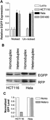
MMR+ cell lines showed significantly higher relative EGFP expression than MMR cell lines. (A) Nicked or un-nicked homo- or heteroduplex (with a G–G mismatch) plasmids were transfected into the cell lines depicted. Percentage of green cells and their mean intensity from each transfection were obtained from flow cytometry, and relative EGFP expression was calculated as described in Materials and Methods. Each column represents mean ± SEM from five independent measurements for HCT116 and HeLa, and from three independent measurements for LoVo and SW480. (B) HCT116 and HeLa cells were co-transfected with nicked homoduplex or heteroduplex EGFP plasmid and RFP plasmid. Twenty-four hours later, the cells were harvested and western blot was done as described in Materials and Methods. (C) The density of EGFP bands was measured with Image-Pro Plus software (Media Cybernetics), normalized with that of the corresponding RFP bands, and plotted in an arbitrary unit.
Figure 5
Figure 5
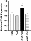
DNA ligase IIIα deficient cell line, CHO EM9, showed higher relative EGFP expression. Nicked homo- and heteroduplex plasmids were transfected into the cell lines depicted in the figure. CHO EM 9 was derived from the parental cell AA8, which is ligase IIIα-proficient. Relative EGFP expression was presented as mean ± SEM from three independent experiments. The asterisk indicates significant difference (P ≤ 0.05) from other mean values with student t tests.
Figure 6
Figure 6
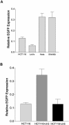
Relative EGFP expression from a T–G mismatch and the effect of chromosome 3 complementation on MMR in HCT116 cell. (A) The same amount of un-nicked heteroduplex (with a T–G mismatch), homoduplex, and negative control DNA were separately transfected into the depicted cell lines. Percentage of green cells and their mean intensity from each transfection were obtained from flow cytometry, and relative EGFP expression was calculated as described in Materials and Methods. Each column represents mean ± SEM from seven independent measurements for HCT116, HeLa and SW480, and six independent measurements for LoVo. According to Newman–Keuls multiple comparison test, the MMR efficiencies of HCT116 and LoVo are significantly different from those of HeLa or SW480 (P < 0.001), whereas the MMR efficiency between the two MMR- cell lines or between the two MMR+ cell lines is not significantly different from each other (P > 0.05). (B) Comparison of T–G MMR in HCT116, HCT116+ch3 and HCT116+ch5 cells. The relative EGFP expression in HCT116 is 0.13 ± 0.01 from 7 independent measurements. The relative EGFP expression in HCT116 + ch3 and HCT116 + ch5 is respectively 0.35 ± 0.05 and 0.13 ± 0.04 from 5 independent measurements. The data are presented as mean ± SEM. According to Newman–Keuls multiple comparison test, the MMR efficiency of HCT116 + ch3 is significantly higher from those of HCT116 and HCT116+ch5 (P < 0.01).
Figure 7
Figure 7
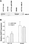
Ectopic expression of hMLH1 increased MMR efficiency in HCT116 cells. (A) Detection of hMLH1 protein with western blotting. HCT116 and SW480 were co-transfected with 1 μg pCMV-MLH1 or 0.6 μg pRC/CMV (control) and the homo- or heteroduplex (with a G–G mismatch) plasmid. Forty-eight hours later, the transfected cells were lifted and analyzed with flow cytometry. Cells recovered from the flow cytometer, along with HCT116+ch3 and SW480 cells, were lysed. Proteins were extracted, separated on 8% SDS-PAGE, and immunoblotted by a hMLH1 antibody. (B) Effect of hMLH1 transfection on G–G MMR in HCT116 and SW480 cells. Relative EGFP expression was calculated after flow cytometry analysis as described in the Materials and Methods. According to student t-test, HCT116 transfected with hMLH1 expression plasmid has a significant higher (P < 0.02) MMR efficiency (0.43 ± 0.05) than HCT116 transfected with pRC/CMV (0.26 ± 0.05). Each column represents mean ± SEM of three independent measurements.

Similar articles

Cited by

References

    1. Modrich P. and Lahue,R. (1996) Mismatch repair in replication fidelity, genetic recombination, and cancer biology. Annu. Rev. Biochem., 65, 101–133. - PubMed
    1. Fishel R. (1998) Mismatch repair, molecular switches, and signal transduction. Genes Dev., 12, 2096–2101. - PubMed
    1. Umar A. and Kunkel,T.A. (1996) DNA-replication fidelity, mismatch repair and genome instability in cancer cells. Eur. J. Biochem., 238, 297–307. - PubMed
    1. Kolodner R.D. and Marsischky,G.T. (1999) Eukaryotic DNA mismatch repair. Curr. Opin. Genet. Dev., 9, 89–96. - PubMed
    1. Modrich P. (1997) Strand-specific mismatch repair in mammalian cells. J. Biol. Chem., 272, 24727–24730. - PubMed

Publication types

MeSH terms