

Review
doi: 10.1073/pnas.0403600101.
Epub 2004 Jul 12.
Discovery of antivirals against smallpox
Affiliations
- PMID: 15249657
- PMCID: PMC509180
- DOI: 10.1073/pnas.0403600101
Item in Clipboard
Review
Discovery of antivirals against smallpox
Proc Natl Acad Sci U S A.
.
No abstract available
Figures
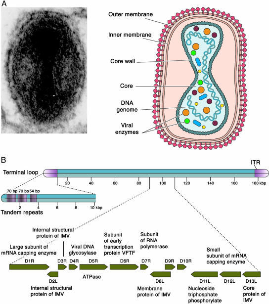
Vaccinia virus, a representative poxvirus: virion structure (A) and genome organization with an expanded view of the HindIII D restriction enzyme fragment (B). The presence of an inner membrane in the IMV form of the virion shown in A is controversial. See Poxvirus replication cycle for a detailed description. [Reproduced with permission from ref. (Copyright 2003, ASM Press).]
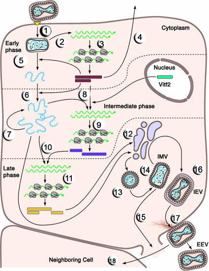
The single-cell reproductive cycle of vaccinia virus. The entry and replication of an EEV are illustrated. RNA molecules are green. See Poxvirus replication cycle for a detailed description of each illustrated step. [Reproduced with permission from ref. (Copyright 2003, ASM Press).]
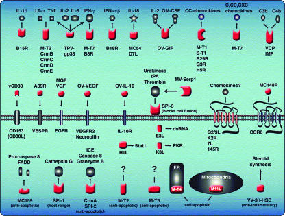
Schematic representation of selected poxvirus immunomodulators and regulators of apoptosis. Proteins shown in red represent poxvirus proteins; host proteins are shown in black and gray. Secreted poxvirus viroceptor proteins (top row) function as soluble or cell surface decoys that bind host-cell cytokines or chemokines in the cell exterior. Poxvirus virokines also are secreted; they function as agonistic or antagonistic ligands for host cellular receptors (middle row). A number of poxvirus proteins function inside the cell to modulate apoptosis, cytokine processing, and host range (red proteins in cell interior). [Reproduced with permission from ref. (Copyright 2003, Annual Reviews, www.annualreviews.org ).]
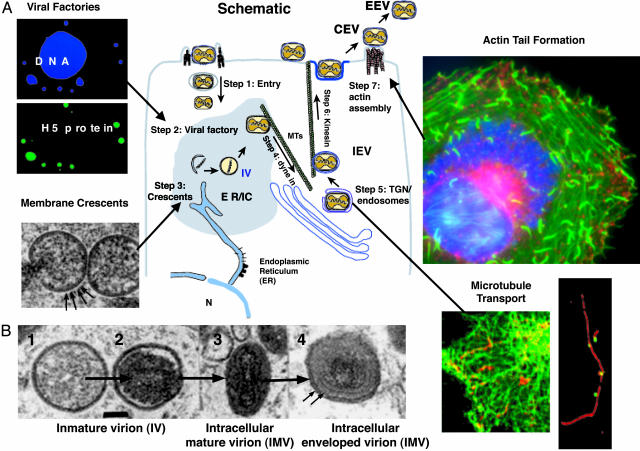
Major questions in the cell biology of poxvirus infection. (A Center) The schematic portrays some events in vaccinia infection and morphogenesis. The specific steps and unanswered questions are numbered and discussed in the text. Stages of virus maturation shown are immature virion (IV), intracellular mature virion (IMV), intracellular enveloped virion (IEV), cell-associated enveloped virion (CEV), and extracellular enveloped virion (EEV). Images surrounding the schematic illustrate some of these events. (Upper Left) Fluorescent micrographs of viral factories, which are detected together with the cell nucleus by the DNA binding dye Hoescht (blue) and, more specifically, by antibodies to the viral protein encoded by the H5 gene (green) [Reproduced with permission from ref. (Copyright 2001, American Society for Cell Biology).] (Lower Left) A thin-section electron micrograph of membrane crescents, an early stage in viral assembly. The derivation of the membrane from either the endoplasmic reticulum (ER) or intermediate compartment (IR) is uncertain, although regularly spaced small spikes can be seen on the outer membrane (arrows). [Reproduced with permission from ref. (Copyright 2002, American Society for Microbiology).] (Lower Near Right) Superimposed frames from a time-lapsed video showing microtubule-based movement of vaccinia virus that appears as red streaks along GFP-labeled microtubule tracks (green). [Reproduced with permission from ref. (Copyright 2001, Nature Publishing Group, www.nature.com ).] (Lower Far Right) Viral particles (green) remain stably associated with microtubules (red) even after extensive extraction of infected cells. [Reproduced with permission from ref. (Copyright 2002, Society for General Microbiology).] (Upper Right) A fluorescent micrograph of actin tail formation (green) juxtaposed to and triggered by cell-surface associated CEV (red), which function to propel the virus particle away from the cell and/or into adjacent cells. 4′,6-Diamidino-2-phenylindole (DAPI) staining (blue) reveals a large viral factory adjacent to the cell nucleus (Photograph courtesy of Tim Newsome and Michael Way). (B) Thin-section electron micrographs of sequential stages of viral maturation. [Reproduced with permission from ref. (Copyright 2001, American Society for Microbiology).]
Comment in
-
Harnessing new science is vital for biodefense and global health.Proc Natl Acad Sci U S A. 2004 Aug 3;101(31):11177. doi: 10.1073/pnas.0404433101. Epub 2004 Jul 12. Proc Natl Acad Sci U S A. 2004. PMID: 15249671 Free PMC article. No abstract available.
References
-
- Institute of Medicine (1999) Assessment of Future Scientific Needs for Variola Virus, ed. Briere, R. (Natl. Acad. Press, Washington DC).
-
- Lane, H. C., Montagne, J. L. & Fauci, A. S. (2001) Nat. Med. 7, 1271–1273. - PubMed
-
- White House (December 13, 2002) Protecting Americans: Smallpox Vaccination Program, press release, www.whitehouse.gov/news/releases/2002/12/20021213-1.html.
-
- Flint, S. J., Enquist, L. W., Racaniello, V. R. & Skalka, A. M. (2003) Principles of Virology: Molecular Biology, Pathogenesis, and Control of Animal Viruses (ASM Press, Washington DC), 2nd Ed.
-
- Moss, B. (2001) in Fields Virology, eds. Knipe, D. M. & Howley, P. M. (Lippincott Williams & Wilkins, Philadelphia), pp. 2849–2883.
Publication types
MeSH terms
Substances
LinkOut - more resources
Full Text Sources
Other Literature Sources
Medical
