. 2004 Jul 20;101(29):10614-9.
doi: 10.1073/pnas.0404146101. Epub 2004 Jul 12.

Drs2p-coupled aminophospholipid translocase activity in yeast Golgi membranes and relationship to in vivo function

Affiliations

Drs2p-coupled aminophospholipid translocase activity in yeast Golgi membranes and relationship to in vivo function

Paramasivam Natarajan et al. Proc Natl Acad Sci U S A. .

Abstract

Aminophospholipid translocases (APLTs) are defined primarily by their ability to flip fluorescent or spin-labeled derivatives of phosphatidylserine (PS) and phosphatidylethanolamine (PE) from the external leaflet of a membrane bilayer to the cytosolic leaflet and are thought to establish phospholipid asymmetry in biological membranes. The identities of APLTs remain unknown, although candidate proteins include the Drs2p/ATPase II subfamily of P-type ATPases. Drs2p from budding yeast localizes to the trans-Golgi network (TGN), and here we show that this membrane contains an ATP-dependent APLT that flips 7-nitro-2-1,3-benzoxadiazol-4-yl (NBD) PS and PE derivatives from the luminal to the cytosolic leaflet. To assess the contribution of Drs2p to this activity, TGN membranes were prepared from strains harboring WT or temperature-sensitive alleles of DRS2 and null alleles of three other potential APLT genes (DNF1, DNF2, and DNF3). Assay of these membranes indicated that Drs2p was required for the ATP-dependent translocation of NBD-PS, whereas no active translocation of NBD-PE or NBD-phosphatidylcholine was detected. The specificity of Drs2p for NBD-PS suggested that translocation of PS would be required for the function of Drs2p in protein transport from the TGN. However, cho1 yeast strains that are unable to synthesize PS do not phenocopy drs2 but instead transport proteins normally via the secretory pathway. In addition, a drs2 cho1 double mutant retains drs2 transport defects. Therefore, whereas NBD-PS is a preferred substrate for Drs2p in vitro, endogenous PS is not an obligatory substrate in vivo for the role Drs2p plays in protein transport.


Figures

Fig. 4.
Fig. 4.
Drs2p- and ATP-dependent translocation of NBD-PS from the luminal to the cytosolic leaflet of the membrane. (A) NBD-PS was incorporated into the cytosolic leaflet of TGN membranes from DRS2 dnf1,2,3Δ and drs2-ts dnf1,2,3Δ strains in the presence or absence of hydroxylamine (HA) and incubated for 2 h at 37°C in the absence of ATP. ATP was then added, and the samples were incubated an additional 2 h at 37°C. HA inhibits a PS decarboxylase activity responsible for converting a portion of the NBD-PS to NBD-PE during these incubations. (B and C) The Drs2p-dependent APLT activity requires Mg2+ but is not coupled to other major ions in the assay buffer. (B) The NBD-PS translocation assay was carried out with WT membranes and ATP in the presence or absence of EDTA. (C) TGN membranes from WT cells were isolated and incubated with NBD-PS by using buffers containing K+ and OAc in place of Na+ and Cl. Translocation kinetics were compared with membranes prepared normally (Na+Cl with or without ATP).
Fig. 1.
Fig. 1.
TGN membrane preparations used for translocation assays contain Drs2p. (A) TGN samples (10 μg each) from WT (BY4742), drs2Δ (ZHY615M2D), DRS2 dnf1,2,3Δ (ZHY409), and drs2-ts dnf1,2,3Δ (ZHY410–3A) strains were subjected to SDS/PAGE and immunoblotted for Drs2p. (B) The specific activity of Kex2p, a marker for the TGN, was determined for each membrane preparation. The Kex2p specific activity was nearly identical for the DRS2 dnf1,2,3Δ and drs2-ts dnf1,2,3Δ isogenic pair.
Fig. 2.
Fig. 2.
NBD phospholipid translocation kinetics across Golgi membranes from WT cells. NBD-PS (A), NBD-PE (B), or NBD-PC (C) was incorporated into the cytosolic leaflet of TGN or early Golgi membranes on ice, which were then incubated for up to 4 h at 37°C with or without ATP. At time points indicated, the NBD phospholipid remaining in the cytosolic leaflet was extracted with fatty-acid-free BSA as described in Materials and Methods and is presented as the percentage of total membrane-associated NBD phospholipid. These data suggest that an APLT is present in the TGN but not in the early Golgi membranes. (Left) TGN for all panels. (Right) Early Golgi for all panels.
Fig. 3.
Fig. 3.
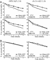
Drs2p function is required for ATP-dependent NBD-PS translocation across the TGN membrane. NBD-PS (A) or NBD-PE (B) was incorporated into the cytosolic leaflet of TGN membranes from DRS2 dnf1,2,3Δ or drs2-ts dnf1,2,3Δ strains, which were then incubated at 27°C(A Upper) or 37°C (A Lower and B). Membranes with WT Drs2p displayed an APLT activity at both temperatures, whereas membranes containing the Drs2-ts protein were temperature-sensitive in this activity. (Left) DRS2 dnf1,2,3Δ for both panels. (Right) drs2-ts dnf1,2,3Δ for both panels.
Fig. 5.
Fig. 5.
Protein transport and processing in the secretory pathway is normal in PS-deficient (cho1) yeast. WT (BY4742), cho1 (JWY2102), and drs2 (ZHY615M2D) were grown at 30°C and labeled for 5 min, and aliquots were removed at the chase times indicated. These strains also were shifted to 15°C for 1 h, labeled for 15 min, and chased for the times indicated. CPY (A) and α-factor (B) were recovered from each sample by immunoprecipitation and subjected to SDS/PAGE as described in ref. .
Fig. 6.
Fig. 6.
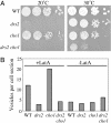
Drs2p function is required for growth at low temperature and transport vesicle formation in cells lacking PS. (A) Growth of WT (BY4742), cho1 (JWY2102), drs2 (ZHY615M2D), and drs2 cho1 (JWY2203) strains on rich medium at 20°C and 30°C. (B) The strains listed above were treated with 0.2 mM LatA for 30 min at 30°C and processed for electron microscopy as described in refs. and . Vesicles were counted, and averages from two experiments are shown.

References

    1. Pomorski, T., Holthuis, J. C., Herrmann, A. & van Meer, G. (2004) J. Cell Sci. 117, 805–813. - PubMed
    1. Williamson, P. & Schlegel, R. A. (1994) Mol. Membr. Biol. 11, 199–216. - PubMed
    1. Seigneuret, M. & Devaux, P. F. (1984) Proc. Natl. Acad. Sci. USA 81, 3751–3755. - PMC - PubMed
    1. Auland, M. E., Roufogalis, B. D., Devaux, P. F. & Zachowski, A. (1994) Proc. Natl. Acad. Sci. USA 91, 10938–10942. - PMC - PubMed
    1. Zachowski, A., Henry, J. P. & Devaux, P. F. (1989) Nature 340, 75–76. - PubMed

Publication types

MeSH terms