. 2004 Jul 20;101(29):10786-91.
doi: 10.1073/pnas.0402876101. Epub 2004 Jul 12.

Trim5alpha protein restricts both HIV-1 and murine leukemia virus

Affiliations

Trim5alpha protein restricts both HIV-1 and murine leukemia virus

Melvyn W Yap et al. Proc Natl Acad Sci U S A. .

Abstract

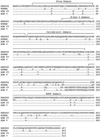
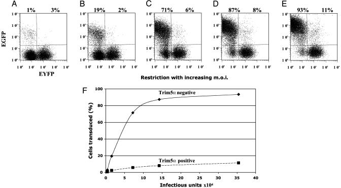
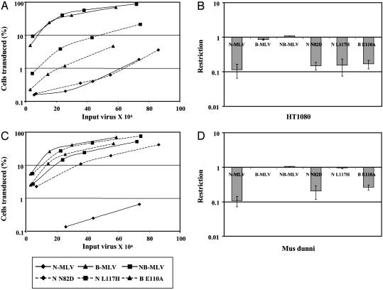
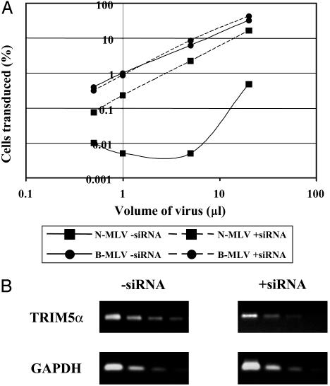
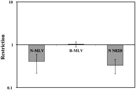
Replication of HIV-1 and N-tropic murine leukemia virus (N-MLV) is restricted in a number of different primate cells. In some cell lines, cross-saturation experiments suggest that the two viruses are interacting with the same restriction factor. Recently, Trim5alpha protein from rhesus monkey was found to restrict HIV-1. We have confirmed this result and have shown that Trim5alpha from two African green monkey cell lines, Vero and CV-1, also restricts HIV-1. In addition, we show that human, rhesus, and African green monkey Trim5alpha can restrict N-MLV. By using a panel of MLV capsid mutants, subtle differences in the anti-MLV activity were identified among the different primate Trim5alpha cDNAs. Trim1 isolated from humans and green monkeys was also found to restrict N-MLV. We hypothesize that the Trim family of proteins plays a widespread role in innate immunity to viral infection.


Figures

Fig. 1.
Fig. 1.
Comparison of amino acid sequence of Trim5α from different primates. Predicted amino acid sequences of the Trim5α protein from human (TE671), rhesus monkey (LLCMK2), AGM-V (Vero), and AGM-CV (CV-1). The positions of the motifs characteristic of Trim proteins are indicated. The sequences of the first eight and last six amino acids were determined by the (human) primers used for amplification.
Fig. 2.
Fig. 2.
Titration of HIV-1 in HT1080 cells transduced with Trim5α from rhesus monkey. (A–E) The FACS profiles of cell populations challenged with increasing amounts of HIV-1 vector carrying the EGFP marker. (A) A total of 4 × 10³ infectious units. (B) A total of 16 × 103 infectious units. (C) A total of 70 × 103 infectious units. (D) A total of 140 × 103 infectious units. (E) A total of 350 × 103 infectious units. Enhanced yellow fluorescent protein (EYFP) identifies Trim5α transduced cells, whereas EGFP follows HIV-1 infection. The percentages of HIV-1 infected cells without and with Trim5α are shown above the profiles. m.o.i., multiplicity of infection. (F) The percentage of infected cells from A–E as a function of the amount of added HIV-1. Data shown are from one typical experiment.
Fig. 3.
Fig. 3.
Restriction of MLV by Trim5α. The graphs on the left show titration curves for a panel of MLVs on TE671 (A) and Vero (C) cell lines. Cells were infected with increasing amounts of viruses (previously titrated on M. dunni cells). Viruses tested were N-MLV (♦, solid line), B-MLV (▴, solid line), NB-MLV (▪, solid line), N-N82D (♦, dashed line), N-L117H (▪, dashed line), and B-E110A (▴, dashed line). The titrations come from one typical experiment. The plots in the right column show the fold reduction (percentage of Trim5α+ cells infected/percentage of Trim5α- cells infected) in MLV titer as measured by the two-color FACS assay upon introduction of human (B) and AGM (D) Trim5α into M. dunni cells. Means and SD from three experiments are shown.
Fig. 4.
Fig. 4.
Effects of Trim5α siRNA on restriction in primate cells. (A) Titration curves of MLV and HIV-1. ▪, TE671: N-MLV; ▪, B-MLV. Continuous lines show titers in the absence of siRNA and broken lines show titers in the presence of siRNA. (B) PCR of 5-fold serial dilutions of Trim5α cDNA isolated from the cells in A. GAPDH was used as a negative control. Data from one representative experiment are shown.
Fig. 5.
Fig. 5.
Restriction of MLV by Trim1. The graph shows the fold reduction (percentage of Trim1+ cells infected/percentage of Trim1- cells infected) in MLV titer as measured by the two-color FACS assay upon introduction of Vero Trim1 into M. dunni cells. Means and SD from three experiments are shown.

Comment in

References

    1. Boeke, J. D. & Stoye, J. P. (1997) in Retroviruses, eds. Coffin, J. M., Hughes, S. H. & Varmus, H. E. (Cold Spring Harbor Lab. Press, Plainview, NY), pp. 343-435. - PubMed
    1. Goodchild, N. L., Wilkinson, D. A. & Mager, D. L. (1993) Virology 196, 778-788. - PubMed
    1. Johnson, W. E. & Coffin, J. M. (1999) Proc. Natl. Acad. Sci. USA 96, 10254-10260. - PMC - PubMed
    1. Belshaw, R., Pereira, V., Katzourakis, A., Paces, J., Burt, A. & Tristem, M. (2004) Proc. Natl. Acad. Sci. USA 101, 4894-4899. - PMC - PubMed
    1. Hasenkrug, K. J. & Chesebro, B. (1997) Proc. Natl. Acad. Sci. USA 94, 7811-7816. - PMC - PubMed

Publication types

MeSH terms