. 2004 Jul 13;32(12):e104.
doi: 10.1093/nar/gnh097.

ChIP Display: novel method for identification of genomic targets of transcription factors

Affiliations

ChIP Display: novel method for identification of genomic targets of transcription factors

Artem Barski et al. Nucleic Acids Res. .

Abstract

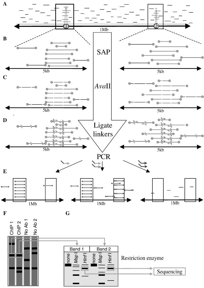
Novel protein-DNA interactions in mammalian cells are traditionally discovered in the course of promoter studies. The genomic era presents opportunities for the reverse; namely, the discovery of novel target genes for transcription factors of interest. Chromatin immunoprecipitation (ChIP) is typically used to test whether a protein binds to a candidate promoter in living cells. We developed a new method, ChIP Display (CD), which allows genome-wide unbiased identification of target genes occupied by transcription factors of interest. Initial CD experiments pursuing target genes for RUNX2, an osteoblast master transcription factor, have already resulted in the identification of four genes that had never been reported as targets of RUNX2. One of them, Osbpl8, was subjected to mRNA and promoter-reporter analyses, which provided functional proof for its regulation by RUNX2. CD will help to assemble the puzzle of interactions between transcription factors and the genome.


Figures

Figure 1
Figure 1
Principles of ChIP Display. (A) Precipitated DNA fragments (green—specific; black—non-specific) are aligned with the genome. Graph (red) shows the representation of each nucleotide in the immunoprecipitate. The area shown contains two hypothetical target genes (#1 and #2). (B) Magnification of the two regions of interest from (A). DNA fragments are treated with shrimp alkaline phosphatase (SAP) to prevent linker ligation to DNA ends generated during sonication. Red circles, dephosphrylated end. (C) DNA is digested with AvaII. (D) Linkers are ligated to the ends of AvaII fragments. Also shown are the nested primers that can amplify targets #1 and #2. (E) PCR products amplified in three reactions: left—target #1 is amplified in a reaction with a single primer (family X in Figure 2B); middle—target #2 is amplified with two different primers (family V in Figure 2B); note that target #1 is amplified here again; right—most of the PCR reactions will amplify neither target #1 nor target #2. (F) Schematic illustration of PAGE of PCR products from two ChIPs and two mock ChIPs (no Ab) belonging to one CD family. (G) Each of the two co-migrating bands from (F) is excised, reamplified and restriction-analyzed with four cutters. Major co-migrating restriction products are then isolated and directly sequenced.
Figure 2
Figure 2
CD families. (A) Design of linkers and nested primers. Positions +1 to +6 are defined at the top. Nucleotides at positions +3 and +6 (highlighted) were used to segregate AvaII fragments into families. Linkers contain a C at position +1 to destroy the AvaII sites and a 1:1 A:T mixture (W) at position +3. In addition, this panel shows one of the eight PCR nested primers, the one with T at position +3 and C at position +6. (B) The eight nested primers produce 36 combinations for PCR. White square—a single primer. Shaded square—two different primers.
Figure 3
Figure 3
CD identifies a novel RUNX2 target gene. (A) Three independent RUNX2 chromatin immunoprecipitates (ChIP) and three no-antibody controls were subjected to CD procedure using a single nested primer (with T and C at positions +3 and +6, respectively; see Figure 1). Amplification products were resolved in 6% polyacrylamide gel and visualized by ethidium bromide staining. The three bands common to all three ChIPs (boxed) were excised. (B) The DNA eluted from each of the bands in (A) was reamplified, digested with either HinfI or MspI, and subjected to gel electrophoresis. Major restriction products were sequenced and all mapped to the vicinity of the Osbpl8 start site. (C) Schematic illustration of a 3 kb genomic region (horizontal line) centered around the Osbpl8 transcription start site. The first exon is represented by the white box. Black box (‘hit’) denotes the AvaII fragment that was amplified during the CD procedure. The hit is located inside a CpG island (striped box) but does not overlap with repetitive sequences (gray boxes). Ten putative RUNX2 binding sites are shown at the top as vertical bars perpendicular to the horizontal line. The darker bars represent those RUNX2 sites that were confirmed by EMSA (Figure 3A). The −751/+12 and +263/+996 fragments, which were cloned into reporter vectors, are shown at the bottom.
Figure 4
Figure 4
Validation of CD hits. Association of RUNX2 with the Osbpl8 gene (O), as well as the Runx3 gene (R), was tested using conventional ChIP assay. Fragments of Osbpl8 (O) and Runx3 (R) genes were PCR amplified with promoter-specific primers listed in Supplementary Table 1. Collagen alpha1(I) (8) (C) and insulin (I) were used as positive and negative controls, respectively. Genomic DNA was used to demonstrate dynamic range of PCR amplification.
Figure 5
Figure 5
Osbpl8 expression is regulated by RUNX2. (A) Osbpl8 transcription start site is surrounded by RUNX2-binding elements. MC3T3-E1 cell extract was subjected to EMSA with 20 bp oligonucleotide probes centered around putative RUNX2 binding sites located at the given positions relative to the Osbpl8 transcription start site. Positive control was the osteoblast-specific element 2 probe (OSE2) from the osteocalcin gene promoter (32). The RUNX2 complex was either competed (C) with unlabeled OSE2 oligonucleotide or supershifted with anti RUNX2 antibodies (Ab). (B) Osbpl8 gene regulatory sequences surrounding the transcription start site confer RUNX2 responsiveness. ST2 marrow stroma-derived cells were transiently transfected with 3 μg of the indicated reporter plasmid and 0.5 μg of the indicated expression vector. Reporter plasmid containing the osteocalcin promoter, a classical RUNX2 target, was used as a positive control (32). Luciferase activity with the empty vector was set at 100 (raw values were 26, 2532, 748 and 838 for -147-OC-luc, -751-osbpl8-luc, osbpl8-intr-luc and TK-luc, respectively). Data are Mean ± SEM (n = 3). (C) Osbpl8 gene expression parallels development of the osteoblast phenotype. ST-2 cells were treated at confluency (day 0) with 300 ng/ml BMP2 to induce osteoblast differentiation. RNA was extracted on the indicated days for RT–PCR analysis of Osbpl8 (O8), osteocalcin (OC) and cyclophylin (CP) mRNAs.

Similar articles

Cited by

References

    1. Liang P. and Pardee,A.B. (1992) Differential display of eukaryotic messenger RNA by means of the polymerase chain reaction. Science, 257, 967–971. - PubMed
    1. Weinmann A.S. and Farnham,P.J. (2002) Identification of unknown target genes of human transcription factors using chromatin immunoprecipitation. Methods, 26, 37–47. - PubMed
    1. Bigler J. and Eisenman,R.N. (1994) Isolation of a thyroid hormone-responsive gene by immunoprecipitation of thyroid hormone receptor–DNA complexes. Mol. Cell. Biol., 14, 7621–7632. - PMC - PubMed
    1. Cawley S., Bekiranov,S., Ng,H.H., Kapranov,P., Sekinger,E.A., Kampa,D., Piccolboni,A., Sementchenko,V., Cheng,J., Williams,A.J. et al. (2004) Unbiased mapping of transcription factor binding sites along human chromosomes 21 and 22 points to widespread regulation of noncoding RNAs. Cell, 116, 499–509. - PubMed
    1. Komori T., Yagi,H., Nomura,S., Yamaguchi,A., Sasaki,K., Deguchi,K., Shimizu,Y., Bronson,R.T., Gao,Y.H., Inada,M. et al. (1997) Targeted disruption of Cbfa1 results in a complete lack of bone formation owing to maturational arrest of osteoblasts. Cell, 89, 755–764. - PubMed

Publication types