. 2004 Jul 20;101(29):10810-4.
doi: 10.1073/pnas.0404161101. Epub 2004 Jul 13.

Nitrosative stress linked to sporadic Parkinson's disease: S-nitrosylation of parkin regulates its E3 ubiquitin ligase activity

Affiliations

Nitrosative stress linked to sporadic Parkinson's disease: S-nitrosylation of parkin regulates its E3 ubiquitin ligase activity

Dongdong Yao et al. Proc Natl Acad Sci U S A. .

Erratum in

  • Proc Natl Acad Sci U S A. 2004 Sep 21;101(38):13969

Abstract

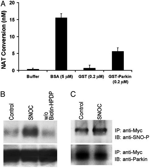
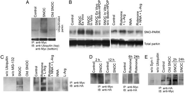
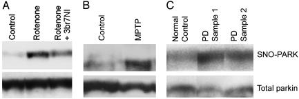
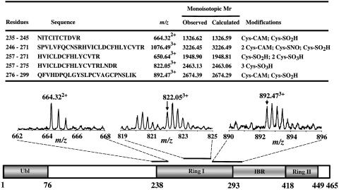
Many hereditary and sporadic neurodegenerative disorders are characterized by the accumulation of aberrant proteins. In sporadic Parkinson's disease, representing the most prevalent movement disorder, oxidative and nitrosative stress are believed to contribute to disease pathogenesis, but the exact molecular basis for protein aggregation remains unclear. In the case of autosomal recessive-juvenile Parkinsonism, mutation in the E3 ubiquitin ligase protein parkin is linked to death of dopaminergic neurons. Here we show both in vitro and in vivo that nitrosative stress leads to S-nitrosylation of wild-type parkin and, initially, to a dramatic increase followed by a decrease in the E3 ligase-ubiquitin-proteasome degradative pathway. The initial increase in parkin's E3 ubiquitin ligase activity leads to autoubiquitination of parkin and subsequent inhibition of its activity, which would impair ubiquitination and clearance of parkin substrates. These findings may thus provide a molecular link between free radical toxicity and protein accumulation in sporadic Parkinson's disease.


Figures

Fig. 1.
Fig. 1.
S-nitrosylation of parkin in vitro. (A) Recombinant protein GST-parkin (0.2μM) was incubated with SNOC (200μM) for 30 min at room temperature (RT). The SNO-PARK thus generated was assessed by release of NO, causing the conversion of 2,3-diaminonaphthalene to the fluorescent compound NAT. GST protein alone was used as negative control. BSA (5 μM) was used as positive control (25). SNOC itself quickly decayed and thus resulted in insignificant S-nitrosothiol readings in this assay. (B)(Upper) Cell lysates from human neuroblastoma SH-SY5Y cells overexpressing myc-tagged parkin were incubated with 200 μM SNOC at RT. Control samples were subjected to decayed SNOC under the same conditions. Thirty minutes after SNOC exposure, SNO-PARK was detected by the biotin-switch assay (15). w/o Biotin-HPDP represents the control without biotin linker [N-[6-biotinamido)hexyl]-1′-(2′pyridyldithio) propionamide] (Lower) Total parkin in cell lysates identified by immunoblot. (C) (Upper) Cell lysates were immunoprecipitated with anti-myc antibody. The immunoprecipitates were incubated with or without SNOC and subjected to SDS/PAGE Western analysis under nonreducing conditions. The membrane was blotted with anti-S-nitrosylated protein antibody to identify nitrosylated parkin and revealed an ≈2-fold increase after SNOC exposure by densitometry. (Lower) Same blot reprobed with anti-parkin antibody to verify equal loading.
Fig. 2.
Fig. 2.
Regulation of parkin E3 ubiquitin ligase activity by S-nitrosylation. (A) In vitro S-nitrosylation of parkin up-regulates its E3 ligase activity. (Upper) Lysates from parkin-overexpressing SH-SY5Y cells were immunoprecipitated with anti-myc antibody. The immunoprecipitates were incubated with SNOC (200 μM) and then subjected to in vitro ubiquitination reaction. SNOC increased the autoubiquitination of parkin, detected by anti-ubiquitin antibody. (Lower) Parkin loading control. (B) S-nitrosylation of parkin after SNOC or rotenone exposure. (Upper) Cell lysates from SH-SY5Y cells that had been exposed to SNOC (SNOC Ex Vivo) or rotenone (1 μM for 6 h) were subjected to the biotin-switch assay to detect SNO-PARK. Also, cell lysates were exposed to SNOC directly (SNOC In Vitro). Rotenone exposure was supplemented with l-arginine (labeled l-Arg) as a NOS substrate. Both SNOC and rotenone increased SNO-PARK in these assays. Nω-nitro-l-arginine (NNA) was used as a NOS inhibitor and suppressed SNO-PARK formation. (Lower) Parkin loading control. (C) Ex vivo S-nitrosylation of parkin up-regulates its E3 ligase activity. SH-SY5Y cells were transfected with HA-tagged ubiquitin and exposed to SNOC or rotenone to nitrosylate parkin. Cell lysates were then subjected to immunoprecipitation with anti-myc followed by Western blot analysis with anti-HA to detect autoubiquitinated parkin. NNA prevented the rotenone-induced increase in E3 ligase activity. (D) Effect of NO donor SNOC and rotenone on parkin E3 activity at different times. SH-SY5Y cells were exposed to SNOC or rotenone. At early time points (2 h after SNOC or 6 h after rotenone), parkin autoubiquitination increased compared with control but decreased several hours later. (E) Effect of NO donor on ubiquitination of synphilin-1 (Syn-1) by parkin. SH-SY5Y cells were cotransfected with HA-synphilin-1 and myc-ubiquitin and exposed to SNOC. Synphilin-1 ubiquitination increased 2 h after SNOC exposure but decreased 24 h later. Old SNOC, decayed SNOC.
Fig. 3.
Fig. 3.
S-nitrosylation of parkin in rodent models and human PD brain. (A) Vehicle (control), rotenone, or rotenone plus the relatively specific neuronal NO synthase inhibitor 3-bromo-7-nitroindazole (3br7NI) was i.p. injected into Sprague–Dawley rats (26). (Upper) Brain extracts from substantia nigra and striatum were subjected to the biotin-switch assay to detect SNO-PARK. Rotenone resulted in increased SNO-PARK that was partially abrogated by 3br7NI. (Lower) Parkin loading control. (B) SNO-PARK was detected in MPTP-, but not control-, injected mice. (C) SNO-PARK was detected in representative temporal cortex of human brain extracts from two sporadic PD patients with diffuse Lewy bodies but not from control brain by using the biotin-switch assay. In total, we tested four brain samples from PD, two normal controls with no CNS disease, two controls with Alzheimer's disease, and two controls with Huntington's disease (see Fig. 6). Each of the PD brain samples, but none of the control brains, manifests evidence for SNO-PARK formation (P < 0.01 by χ2 test).
Fig. 4.
Fig. 4.
Peptide mass fingerprinting analysis of the modified MS thiol group of cysteine residues within the highly conserved RING I domain of recombinant human parkin exposed to SNOC. (Upper) Quadrupole time-of-flight (Q-TOF) results for RING I domain of recombinant parkin exposed to SNOC. Sequences containing modified cysteines. Carbamidomethyl (CAM) signifies modification by iodoacetamide alkylation to protect free cysteines before trypsin digestion. Cys-SNO indicates S-nitrosothiol formation; Cys-SO2H or Cys-SO3H indicates sulfinic or sulfonic acid derivatization, respectively. Previously, we had shown that nitrosylation can precede further oxidation to these derivatives (14). Five of the seven cysteine residues in the RING I domain underwent modification. Single-letter abbreviations for the amino acid residues are as follows: A, Ala; C, Cys; D, Asp; E, Glu; F, Phe; G, Gly; H, His; I, Ile; K, Lys; L, Leu; M, Met; N, Asn; P, Pro; Q, Gln; R, Arg; S, Ser; T, Thr; V, Val; W, Trp; and Y, Tyr. (Lower) Schematic representation of parkin and sample mass spectra of parkin modifications in RING I domain (bars indicate tryptic fragments containing modified cysteine residues). Ubl, ubiquitin-like domain; IBR, in-between RING domain. For additional mass spectra results on the RING II and IBR domains, see Fig. 7 and Table 1.
Fig. 5.
Fig. 5.
Model of S-nitrosylation of parkin. Ribbon structure of a partial sequence of human parkin showing RING I domain (residues 237Thr to 291Ala). α-Helix shown in red, β-sheet in cyan, cysteine residues in green, histidine in yellow, aspartate in red, and arginine in blue. 241Cys and 260Cys match the nitrosylation consensus motif (13).

References

    1. Mayer, R. J., Lowe, J., Lennox, G., Doherty, F. & Landon, M. (1989) Prog. Clin. Biol. Res. 317, 809-818. - PubMed
    1. Bence, N. F., Sampat, R. M. & Kopito, R. R. (2001) Science 292, 1552-1555. - PubMed
    1. Goldberg, A. L. (2003) Nature 426, 895-899. - PubMed
    1. Perutz, M. F. & Windle, A. H. (2001) Nature 412, 143-144. - PubMed
    1. Yong, V. W., Power, C., Forsyth, P. & Edwards, D. R. (2001) Nat. Rev. Neurosci. 2, 502-511. - PMC - PubMed

Publication types

MeSH terms