Comparative Study
. 2004 Jul 14;24(28):6291-300.
doi: 10.1523/JNEUROSCI.0586-04.2004.

Behavioral state instability in orexin knock-out mice

Affiliations
Comparative Study

Behavioral state instability in orexin knock-out mice

Takatoshi Mochizuki et al. J Neurosci. .

Abstract

Narcolepsy is caused by a lack of orexin (hypocretin), but the physiologic process that underlies the sleepiness of narcolepsy is unknown. Using orexin knock-out (KO) mice as a model of narcolepsy, we critically tested the three leading hypotheses: poor circadian control of sleep and wakefulness, inadequate activation of arousal regions, or abnormal sleep homeostasis. Compared with wild-type (WT) littermates, orexin KO mice had essentially normal amounts of sleep and wake, but wake and non-rapid eye movement (NREM) bouts were very brief, with many more transitions between all behavioral states. In constant darkness, orexin KO mice had normal amplitude and timing of sleep-wake rhythms, providing no evidence for disordered circadian control. When placed in a new, clean cage, both groups of mice remained awake for approximately 45 min, demonstrating that, even in the absence of orexin, fundamental arousal regions can be engaged to produce sustained wakefulness. After depriving mice of sleep for 2-8 hr, orexin KO mice recovered their NREM and rapid eye movement sleep deficits at comparable rates and to the same extent as WT mice, with similar increases in EEG delta power, suggesting that their homeostatic control of sleep is normal. These experiments demonstrate that the fragmented wakefulness of orexin deficiency is not a consequence of abnormal sleep homeostasis, poor circadian control, or defective fundamental arousal systems. Instead, the fragmented behavior of orexin KO mice may be best described as behavioral state instability, with apparently low thresholds to transition between states.


Figures

Figure 1.
Figure 1.
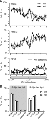
Orexin KO mice have nearly normal amounts of wake and sleep on a 12 hr LD cycle. A, Orexin KO mice have normal hourly amounts of wake and NREM sleep and slightly more REM sleep. Cataplexy occurs almost exclusively during the dark period, especially during times of relatively little REM sleep. B, Across the 12hr dark and light periods, WT and orexin KO mice have similar amounts of NREM and REM sleep. Orexin KO mice have slightly less wake during the dark period, perhaps because of cataplexy during this period. This and all other experiments used seven male orexin KO mice and eight WT male littermates. *p < 0.05; **p < 0.01 compared with WT mice.
Figure 2.
Figure 2.
Orexin KO mice have fragmented wakefulness and sleep on a 12 hr LD cycle. A, Orexin KO mice have very short bouts of wake and NREM sleep. REM bouts are slightly shorter only during the light period, and episodes of cataplexy last approximately as long as REM sleep bouts. B, Orexin KO mice have a greater number of wake and sleep bouts during the light and dark periods. C, During the dark period, most wakefulness in WT mice occurs in bouts longer than 1280 sec (21.3 min), but most wakefulness in orexin KO mice occurs in bouts lasting only 80-1270 sec (1.3-21.2 min). In this time-weighted frequency histogram, the number of bouts of wakefulness has been normalized by the amount of wakefulness occurring over this interval. D, After normalizing for the total number of cycles, orexin KO mice have more short sleep cycles and fewer long cycles during the light period. *p < 0.05; **p < 0.01.
Figure 3.
Figure 3.
Orexin KO mice have more transitions between all behavioral states. The mean number of transitions between states is indicated along the arrows between states and by the thickness of the arrows. All transitions are significantly increased in orexin KO mice, and transitions into cataplexy account for only a small number of the transitions out of wakefulness. *p < 0.05; **p < 0.01.
Figure 4.
Figure 4.
The distribution of EEG power during NREM and REM sleep is similar in WT and orexin KO mice. During cataplexy, the EEG spectra of orexin KO mice is very similar to that seen in REM sleep. The data include all artifact-free sleep and cataplexy epochs during the 12 hr dark period.
Figure 5.
Figure 5.
In constant darkness, orexin KO mice have nearly normal amounts of wakefulness and sleep. A, Orexin KO and WT mice have similar hourly amounts of wake, NREM, and REM sleep. As in the LD condition, most cataplexy occurs during the subjective dark period. B, Across the subjective dark and light periods, orexin KO mice have nearly normal amounts of wake, NREM, and REM sleep, except for slightly less wake during the subjective light period. *p < 0.05.
Figure 6.
Figure 6.
Orexin KO mice have fragmented sleep and wake in constant darkness. A, B, Orexin KO mice have shorter and more frequent bouts of wake and NREM sleep, but these abnormalities are apparent in REM sleep only during the subjective light period. A few episodes of cataplexy occur in the subjective light period. C, During the subjective dark period, most wakefulness in orexin KO mice occurs in short- to mid-length bouts. **p < 0.01.
Figure 7.
Figure 7.
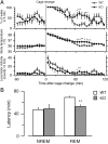
Placing WT and orexin KO in new, clean cages at 1:00 P.M. increases wakefulness, body temperature, and locomotor activity. A, After an initial period of wakefulness, both groups return to sleep after 45 min, but, 60-90 min after cage change, orexin KO mice have more wakefulness and a small rise in body temperature. Orexin KO mice also have a smaller initial increase in locomotor activity. B, After cage change, orexin KO mice remain awake for over 45 min just as seen in WT mice, but they enter REM sleep more rapidly. *p < 0.05; **p < 0.01.
Figure 8.
Figure 8.
Orexin KO mice have nearly normal responses to 8 hr of sleep deprivation. A, Compared with the baseline day (open circles), WT mice have a marked increase in NREM sleep after 8 hr of sleep deprivation (filled circles). WT mice also have rebound REM sleep, primarily during the dark period. B, Orexin knock-out mice have normal rebound of NREM sleep but relatively more rebound REM sleep during the dark period. Although not statistically significant, cataplexy appears to be less frequent after sleep deprivation. Horizontal brackets mark the sleep deprivation (SD) period. *p < 0.05; ** p < 0.01 compared with the baseline day. †p < 0.05; ††p < 0.01 compared with WT mice on the sleep deprivation day.
Figure 9.
Figure 9.
After sleep deprivation, orexin KO mice recover NREM and REM sleep normally, but NREM sleep is still fragmented. A, When compared with the baseline day, both WT and orexin KO mice accumulate a NREM sleep deficit of ∼240 min over the 8 hr of sleep deprivation. Both groups then recover from this deficit at the same rate and to the same extent. B, At baseline, orexin KO mice have shorter bouts of NREM sleep than WT mice. After 8 hr of sleep deprivation, both groups produce longer bouts of NREM sleep, but orexin KO mice still have shorter than normal NREM bouts. *p < 0.05; **p < 0.01.
Figure 10.
Figure 10.
Orexin KO mice have normal, dose-dependent responses to sleep deprivation. A, The amount of rebound NREM and REM sleep in the 4 hr immediately after sleep deprivation increases in proportion to the duration of sleep deprivation. B, In both groups, the latency to enter NREM and REM sleep becomes shorter with increasing durations of sleep deprivation. Latencies are similar between groups, except that orexin KO mice enter REM sleep more rapidly after 8hr of sleep deprivation. C, EEG delta power (0.5-4Hz) during NREM sleep in the 4 hr after sleep deprivation increases with increasing duration of sleep deprivation in both groups. Theta power (4-9Hz) during REM sleep does not change with sleep deprivation. *p < 0.05; **p < 0.01 compared with other durations of sleep deprivation. ††p < 0.01 compared with WT mice.

References

    1. Abrahamson EE, Leak RK, Moore RY (2001) The suprachiasmatic nucleus projects to posterior hypothalamic arousal systems. NeuroReport 12: 435-440. - PubMed
    1. Alam MN, Gong H, Alam T, Jaganath R, McGinty D, Szymusiak R (2002) Sleep-waking discharge patterns of neurons recorded in the rat perifornical lateral hypothalamic area. J Physiol (Lond) 538: 619-631. - PMC - PubMed
    1. Aston-Jones G, Chen S, Zhu Y, Oshinsky ML (2001) A neural circuit for circadian regulation of arousal. Nat Neurosci 4: 732-738. - PubMed
    1. Besset A, Tafti M, Nobile L, Billiard M (1994) Homeostasis and narcolepsy. Sleep 17: S29-S34. - PubMed
    1. Borbély AA, Tobler I (1996) Sleep regulation: relation to photoperiod, sleep duration, waking activity, and torpor. In: Progress in brain research (Buijs RM, Kalsbeek A, Romijn HJ, Pennartz CMA, Nirmiran M, eds), pp 343-348. Amsterdam: Elsevier. - PubMed

Publication types

MeSH terms

LinkOut - more resources