

The amygdala is enlarged in children but not adolescents with autism; the hippocampus is enlarged at all ages
- PMID: 15254095
- PMCID: PMC6729537
- DOI: 10.1523/JNEUROSCI.1297-04.2004
The amygdala is enlarged in children but not adolescents with autism; the hippocampus is enlarged at all ages
Abstract
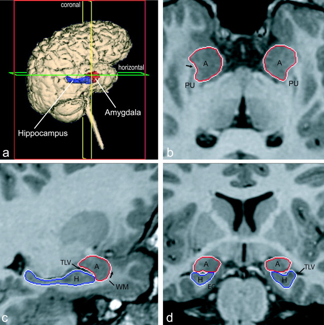
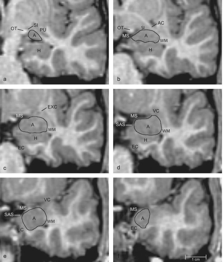
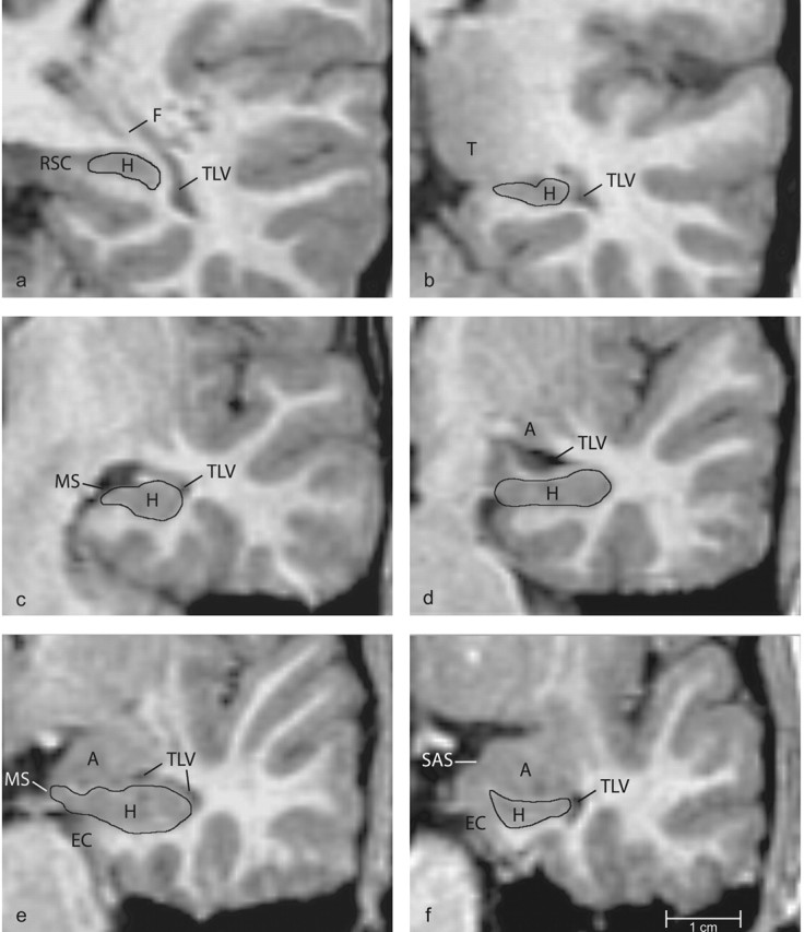
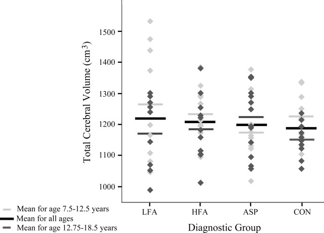
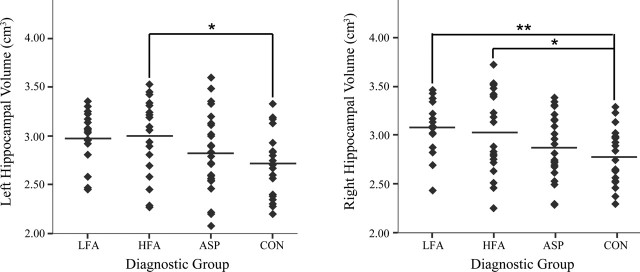
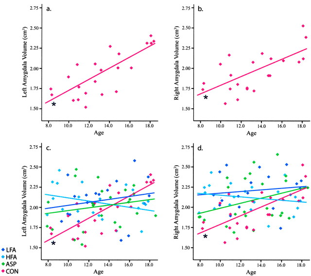
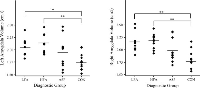
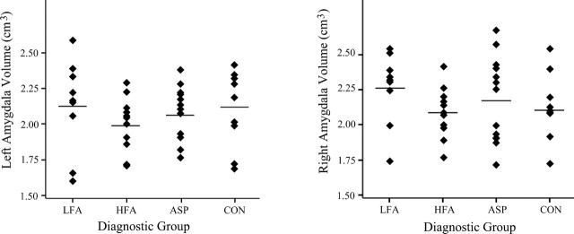
Autism is a neurodevelopmental disorder characterized by impairments in reciprocal social interaction, deficits in verbal and nonverbal communication, and a restricted repertoire of activities or interests. We performed a magnetic resonance imaging study to better define the neuropathology of autistic spectrum disorders. Here we report findings on the amygdala and the hippocampal formation. Borders of the amygdala, hippocampus, and cerebrum were defined, and their volumes were measured in male children (7.5-18.5 years of age) in four diagnostic groups: autism with mental retardation, autism without mental retardation, Asperger syndrome, and age-matched typically developing controls. Although there were no differences between groups in terms of total cerebral volume, children with autism (7.5-12.5 years of age) had larger right and left amygdala volumes than control children. There were no differences in amygdala volume between the adolescent groups (12.75-18.5 years of age). Interestingly, the amygdala in typically developing children increases substantially in volume from 7.5 to 18.5 years of age. Thus, the amygdala in children with autism is initially larger, but does not undergo the age-related increase observed in typically developing children. Children with autism, with and without mental retardation, also had a larger right hippocampal volume than typically developing controls, even after controlling for total cerebral volume. Children with autism but without mental retardation also had a larger left hippocampal volume relative to controls. These cross-sectional findings indicate an abnormal program of early amygdala development in autism and an abnormal pattern of hippocampal development that persists through adolescence. The cause of amygdala and hippocampal abnormalities in autism is currently unknown.
Figures
References
-
- Abell F, Krams M, Ashburner J, Passingham R, Friston K, Frackowiak R, Happe F, Frith C, Frith U (1999) The neuroanatomy of autism: a voxel-based whole brain analysis of structural scans. NeuroReport 10: 1647-1651. - PubMed
-
- Adolphs R (2002) Neural systems for recognizing emotion. Curr Opin Neurobiol 12: 169-177. - PubMed
-
- Adolphs R, Tranel D, Damasio H, Damasio A (1994) Impaired recognition of emotion in facial expressions following bilateral damage to the human amygdala. Nature 372: 669-672. - PubMed
-
- Adolphs R, Tranel D, Damasio AR (1998) The human amygdala in social judgment. Nature 393: 470-474. - PubMed
Publication types
MeSH terms
Grants and funding
- R01 MH050047/MH/NIMH NIH HHS/United States
- P50 CA095817/CA/NCI NIH HHS/United States
- K01 MH001832/MH/NIMH NIH HHS/United States
- HD31715/HD/NICHD NIH HHS/United States
- MH50047/MH/NIMH NIH HHS/United States
- MH01832/MH/NIMH NIH HHS/United States
- R01 MH041479/MH/NIMH NIH HHS/United States
- MH41479/MH/NIMH NIH HHS/United States
- R01 HD031715/HD/NICHD NIH HHS/United States
- R01 NS016980/NS/NINDS NIH HHS/United States
- MH01142/MH/NIMH NIH HHS/United States
- NS16980/NS/NINDS NIH HHS/United States
- R37 MH041479/MH/NIMH NIH HHS/United States
LinkOut - more resources
Full Text Sources