. 2004 Aug;78(15):7874-82.
doi: 10.1128/JVI.78.15.7874-7882.2004.

Adeno-associated virus site-specific integration and AAVS1 disruption

Affiliations

Adeno-associated virus site-specific integration and AAVS1 disruption

Henry Hamilton et al. J Virol. 2004 Aug.

Abstract

Adeno-associated virus (AAV) is a single-stranded DNA virus with a unique biphasic lifestyle consisting of both a productive and a latent phase. Typically, the productive phase requires coinfection with a helper virus, for instance adenovirus, while the latent phase dominates in healthy cells. In the latent state, AAV is found integrated site specifically into the host genome at chromosome 19q13.4 qtr (AAVS1), the only animal virus known to integrate in a defined location. In this study we investigated the latent phase of serotype 2 AAV, focusing on three areas: AAV infection, rescue, and integration efficiency as a function of viral multiplicity of infection (MOI); efficiency of site-specific integration; and disruption of the AAVS1 locus. As expected, increasing the AAV MOI resulted in an increase in the percentage of cells infected, with 80% of cells infected at an MOI of 10. Additional MOI only marginally effected a further increase in percentage of infected cells. In contrast to infection, we found very low levels of integration at MOIs of less than 10. At an MOI of 10, at which 80% of cells are infected, less than 5% of clonal cell lines contained integrated AAV DNA. At an MOI of 100 or greater, however, 35 to 40% of clonal cell lines contained integrated AAV DNA. Integration and the ability to rescue viral genomes were highly correlated. Analysis of integrated AAV indicated that essentially all integrants were AAVS1 site specific. Although maximal integration efficiency approached 40% of clonal cell lines (essentially 50% of infected cells), over 80% of cell lines contained a genomic disruption at the AAVS1 integration locus on chromosome 19 ( approximately 100% of infected cells). Rep expression by itself and in the presence of a plasmid integration substrate was able to mediate this disruption of the AAVS1 site. We further characterized the disruption event and demonstrated that it resulted in amplification of the AAVS1 locus. The data are consistent with a revised model of AAV integration that includes preliminary expansion of a defined region in AAVS1.


Figures

FIG. 1.
FIG. 1.
AAV infection efficiency. HeLa cells were infected with wild-type AAV at various MOIs and coinfected with wild-type adenovirus at an MOI of 10 at 24 h. At 20 h post-adenovirus infection, single-cell suspensions were made as described in Materials and Methods. These were then diluted, filtered onto nylon membranes, and probed with a rep gene fragment to score for productively infected cells. Membranes were then stripped and reprobed with HeLa genomic DNA to score for total cells. A representative filter from a wild-type AAV MOI 100 infection is shown in panel A. Panel B graphs MOIs from 0 to 1,000. Each point represents the average of four individual blots.
FIG. 2.
FIG. 2.
Rescue efficiency in clonal cell lines. HeLa cells were infected with wild-type AAV at specific MOIs and plated out at 24 h to make single-cell clones. Three weeks postinfection, individual cell lines were examined for the presence of latent AAV by a rescue assay. Briefly, cells were seeded in 96-well dishes and infected with wild-type adenovirus at an MOI of 10. At full cytopathic effect (48 to 72 h), cells were lysed by addition of NaOH and EDTA and denatured for 15 min at 68°C. Lysates were filtered onto nylon membranes with a slot blot apparatus and probed for the presence of wild-type AAV DNA. A representative blot from a wild-type AAV MOI 100 infection is shown in panel A. Panel B graphs MOIs from 0 to 1,000. Each point represents a minimum of 36 cell lines screened.
FIG. 3.
FIG. 3.
AAVS1 disruption and AAV DNA persistence. Graph of integration and AAVS1 disruption data from Southern blots of HeLa genomic DNA digested with EcoRI. Each point represents the percentage of clonal cell lines that contained AAVS1 disruptions or integrated AAV at the various MOIs listed.
FIG. 4.
FIG. 4.
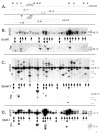
Site-specific integration by AAV. Representative genomic Southern blots showing DNA from HeLa cells infected with wild-type AAV at an MOI of 100. At 6 weeks postinfection, genomic DNA was harvested from clonal cell lines and digested with EcoRI (B), BamHI (C), or ApaLI (D). Duplicate 1% agarose gels were blotted and probed with either Rep or AAVS1, as indicated. A restriction digestion schematic of the AAVS1 site and wild-type AAV-2 is shown in panel A. Size markers are illustrated on the left, as is the position of the wild-type AAVS1 DNA fragment. The AAVS1 and AAV probes are indicated with asterisks. The AAVS1 terminal resolution site integration signal is indicated by shading. Restriction fragment lengths are listed below the DNA schematic. Cell lines containing AAVS1 disruptions or AAVS1 disruptions plus integrated AAV are indicated with single and double arrows, respectively. Note that all integrations occurred in AAVS1-disrupted cell lines.
FIG. 5.
FIG. 5.
AAVS1 region expansion. (A) Genomic Southern blot of randomly selected AAV-infected clonal cell lines probed with the AAVS1 genomic fragment or with a human β-actin probe. HeLa lane, uninfected cells. Class 1 lanes, nondisrupted. Class 2 lanes, disrupted only. Class 3 lanes, disrupted plus integrated. Southern blots were exposed to a phosphorimager cassette, and signal intensities were determined. The average of the experimental sample AAVS1 band intensity or β-actin band intensity was normalized to the value of uninfected HeLa cell AAVS1 band intensity or β-actin, and the data are presented in panel B.
FIG. 6.
FIG. 6.
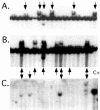
Rep mediated AAVS1 disruption. Representative genomic Southern blots showing DNA from clonal HeLa cell lines transfected with either p78Rep (A) or p78Rep plus a plasmid integration substrate containing the p5IEE-CAT gene (B and C). Panels A and B were probed with AAVS1, and panel C was probed with CAT. Disruptions and integrations are indicated with arrows. C+, positive control genomic DNA obtained from a HeLa-derived cell line containing p5IEE-CAT integrated site specifically into AAVS1 (27).
FIG. 7.
FIG. 7.
Proposed model for Rep-mediated AAVS1 disruption and AAV site-specific integration. (A) Representation of the AAV genome with the inverted terminal repeats hairpinned. A.1, minimum p5 integration element including Rep binding sites (RBS), the transcription factor YY1, and the TATA box. (B) Representation of AAVS1, including the Rep binding site (RBS) and the nicking site for Rep endonuclease activity (trs). Panels B.1 to B.3 diagram Rep binding and replication initiated with host polymerase. Note that Rep is covalently attached to the 5′ end of the nick site. Rep may reinitiate this process several times, perhaps accounting for amplification of AAVS1 (C.2). For integration to occur, a Rep-mediated recombination event between AAVS1 (B.3) and p5 (A.2) results in AAV sequence in the disrupted chromosome 19 locus (C.1).

References

    1. Bartlett, J. S., R. Wilcher, and R. J. Samulski. 2000. Infectious entry pathway of adeno-associated virus and adeno-associated virus vectors. J. Virol. 74:2777-2785. - PMC - PubMed
    1. Beaton, A., P. Palumbo, and K. I. Berns. 1989. Expression from the adeno-associated virus p5 and p19 promoters is negatively regulated in trans by the rep protein. J. Virol. 63:4450-4454. - PMC - PubMed
    1. Berns, K. I., T. C. Pinkerton, G. F. Thomas, and M. D. Hoggan. 1975. Detection of adeno-associated virus (AAV)-specific nucleotide sequences in DNA isolated from latently infected Detroit 6 cells. Virology 68:556-560. - PubMed
    1. Berns, K. I., and R. M. Linden. 1995. The cryptic life style of adeno-associated virus. Bioessays 17:237-245. - PubMed
    1. Berns, K. I. 1996. Parvoviridae: the viruses and their replication, p. 1017-1041. In B. N. Fields (ed.), Fundamental virology, 3rd ed. Lippincott-Raven Publishers, Philadelphia Pa.

Publication types