

Compartmentalization of human immunodeficiency virus type 1 between blood monocytes and CD4+ T cells during infection
- PMID: 15254161
- PMCID: PMC446117
- DOI: 10.1128/JVI.78.15.7883-7893.2004
Compartmentalization of human immunodeficiency virus type 1 between blood monocytes and CD4+ T cells during infection
Abstract
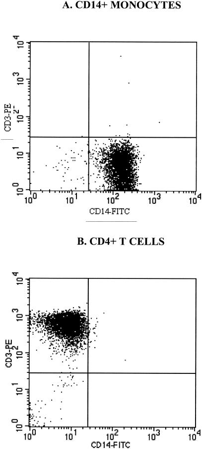
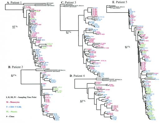
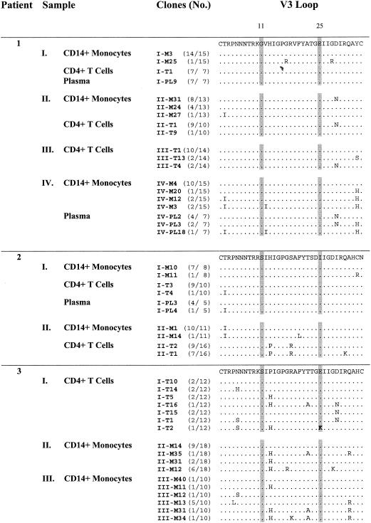
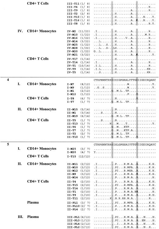
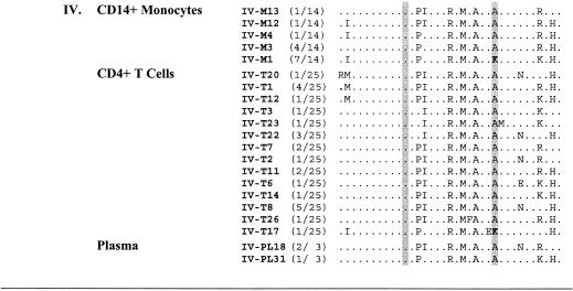
Distinct sequences of human immunodeficiency virus type 1 (HIV-1) have been found between different tissue compartments or subcompartments within a given tissue. Whether such compartmentalization of HIV-1 occurs between different cell populations is still unknown. Here we address this issue by comparing HIV-1 sequences in the second constant region through the fifth hypervariable region (C2 to V5) of the surface envelope glycoprotein (Env) between viruses in purified blood CD14(+) monocytes and CD4(+) T cells obtained longitudinally from five infected patients over a time period ranging from 117 to 3,409 days postseroconversion. Viral populations in both cell types at early infection time points appeared relatively homogeneous. However, later in infections, all five patients showed heterogeneous populations in both CD14(+) monocytes and CD4(+) T cells. Three of the five patients had CD14(+) monocyte populations with significantly more genetic diversity than the CD4(+) T-cell population, while the other two patients had more genetic diversity in CD4(+) T cells. The cellular compartmentalization of HIV-1 between CD14(+) monocytes and CD4(+) T cells was not seen early during infections but was evident at the later time points for all five patients, indicating an association of viral compartmentalization with the time course of HIV-1 infection. The majority of HIV-1 V3 sequences indicated a macrophage-tropic phenotype, while a V3 sequence-predicted T-cell tropic virus was found in the CD4(+) T cells and CD14(+) monocytes of two patients. These findings suggest that HIV-1 in CD14(+) monocytes could disseminate and evolve independently from that in CD4(+) T cells over the course of HIV-1 infection, which may have implications on the development of new therapeutic strategies.
Figures
References
-
- Aquaro, S., R. Calio, E. Balestra, P. Bagnarelli, A. Cenci, A. Bertoli, B. Tavazzi, D. Di Pierro, M. Francesconi, D. Abdelahad, and C. F. Perno. 1998. Clinical implications of HIV dynamics and drug resistance in macrophages. J. Biol. Regul. Homeost. Agents 12:23-27. - PubMed
-
- Aquaro, S., R. Calio, J. Balzarini, M. C. Bellocchi, E. Garaci, and C. F. Perno. 2002. Macrophages and HIV infection: therapeutical approaches toward this strategic virus reservoir. Antivir. Res. 55:209-225. - PubMed
-
- Aquaro, S., C. F. Perno, E. Balestra, J. Balzarini, A. Cenci, M. Francesconi, S. Panti, F. Serra, N. Villani, and R. Calio. 1997. Inhibition of replication of HIV in primary monocyte/macrophages by different antiviral drugs and comparative efficacy in lymphocytes. J. Leukoc. Biol. 62:138-143. - PubMed
-
- Ball, J. K., E. C. Holmes, H. Whitwell, and U. Desselberger. 1994. Genomic variation of human immunodeficiency virus type 1 (HIV-1): molecular analyses of HIV-1 in sequential blood samples and various organs obtained at autopsy. J. Gen. Virol. 75:67-69. - PubMed
Publication types
MeSH terms
Substances
Grants and funding
LinkOut - more resources
Full Text Sources
Medical
Research Materials
Miscellaneous
