. 2004 Aug;78(15):7894-903.
doi: 10.1128/JVI.78.15.7894-7903.2004.

Canine distemper virus and measles virus fusion glycoprotein trimers: partial membrane-proximal ectodomain cleavage enhances function

Affiliations

Canine distemper virus and measles virus fusion glycoprotein trimers: partial membrane-proximal ectodomain cleavage enhances function

Veronika von Messling et al. J Virol. 2004 Aug.

Abstract

The trimeric fusion (F) glycoproteins of morbilliviruses are activated by furin cleavage of the precursor F(0) into the F(1) and F(2) subunits. Here we show that an additional membrane-proximal cleavage occurs and modulates F protein function. We initially observed that the ectodomain of approximately one in three measles virus (MV) F proteins is cleaved proximal to the membrane. Processing occurs after cleavage activation of the precursor F(0) into the F(1) and F(2) subunits, producing F(1a) and F(1b) fragments that are incorporated in viral particles. We also detected the F(1b) fragment, including the transmembrane domain and cytoplasmic tail, in cells expressing the canine distemper virus (CDV) or mumps virus F protein. Six membrane-proximal amino acids are necessary for efficient CDV F(1a/b) cleavage. These six amino acids can be exchanged with the corresponding MV F protein residues of different sequence without compromising function. Thus, structural elements of different sequence are functionally exchangeable. Finally, we showed that the alteration of a block of membrane-proximal amino acids results in diminished fusion activity in the context of a recombinant CDV. We envisage that selective loss of the membrane anchor in the external subunits of circularly arranged F protein trimers may disengage them from pulling the membrane centrifugally, thereby facilitating fusion pore formation.


Figures

FIG. 1.
FIG. 1.
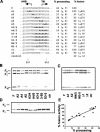
Scheme of the CDV F protein and sequences of the membrane-proximal regions of three paramyxoviral F proteins (A) and Western blot analysis of different paramyxoviral F proteins (B and C). (A, top) Linear drawing of the CDV F protein. This protein is synthesized with a long amino-terminal precursor sequence that is cleaved posttranslationally prior to cleavage activation of the F0 precursor into the disulfide-linked F1 and F2 subunits (53). Hydrophobic regions are indicated by hatched boxes. F protein subunits are labeled as follows: signal peptide (SP), F0 precursor, F1 and F2 subunits, and the F1a and F1b fragments that result from the newly identified cleavage. The disulfide bond (SS) connecting the F1 and F2 subunits is shown below the F protein scheme. The boxes marked HRA and HRB indicate the positions of the corresponding heptad repeats. (Bottom) Alignment of sequences surrounding the putative cleavage regions of CDV, MV, and MuV F proteins. Identical residues are indicated by dots, hydrophobic residues in the first and fourth (a and d) positions of HRB are in bold, and predicted transmembrane domain residues are italicized. (B and C) Characterization of the different F protein forms by Western blot analysis. Proteins were extracted from purified CDV or MV particles (par) or Vero cells transfected (tr) with the CDV, MV, or MuV F protein expression plasmids or infected (inf) with CDV or MV, or control uninfected cells (ctr). Lysates were separated by reducing SDS-PAGE and blotted onto polyvinylidene difluoride membranes. The F proteins were revealed with anti-cytoplasmic tail (B) or anti-F431 (C) serum. The positions of F0, F1, F1a, and F1b are indicated on the left.
FIG. 2.
FIG. 2.
Trypsin digestion of CDV F protein mutants. Western blot analysis of crude membrane preparations of Vero cells infected with CDV or transfected with different F protein expression plasmids was performed. Cells underwent hypotonic lysis prior to pelleting through 250 mM sucrose in TBS. The pellet was resuspended and divided into two equal aliquots, one of which was subjected to trypsin digestion (+), while the other served as a control (−). Lysates were separated by reducing SDS-PAGE (15% acrylamide) and blotted onto polyvinylidene difluoride membranes. The F proteins were revealed with an anti-cytoplasmic tail serum. The positions of F0, F1, and F1b are indicated on the left. Note that trypsin digestion reduced the amount of all F protein forms detected regardless of the mutant analyzed. C, control nontransfected cells; F, Ftryps, and FER, cells transfected with pCG-F, pCG-Ftryps, and pCG-FER, respectively.
FIG. 3.
FIG. 3.
Scheme of the F protein (A), sequence of its membrane-proximal region (B), and initial mapping of the membrane-proximal cleavage site (C). (A) Scheme of the membrane-bound F protein (left) and of the truncated F proteins sF608, sF595, and sF585. The disulfide bond connecting the F1 and F2 subunits is symbolized by a line. The cell membrane is indicated by a gray rectangle, the Flag tag by a white box, and the cleavage site by an arrow. (B) The position of the last residue of different proteins is indicated with an ordinal number and a bent arrow. The hydrophobic a and d residues of HRB are in bold, and predicted transmembrane domain residues are italicized. (C) Western blot analysis of soluble F proteins. Supernatants of Vero cells transfected with the indicated plasmids were subjected to reducing SDS-PAGE (10% acrylamide) and blotted onto polyvinylidene difluoride membranes. Soluble F proteins were detected with a monoclonal antibody directed against the Flag peptide that was added at the truncation site. The positions of F0 and F1 are indicated on the left.
FIG. 4.
FIG. 4.
Sequence, processing, fusion activity, and surface expression of CDV F protein cleavage mutants. (A) Sequence of the region of interest of the original and mutant F proteins, efficacy of their membrane-proximal processing, and fusion activity. The names of the mutant F proteins are indicated on the left. Mutated residues are bold. The end of HRB and the beginning of the transmembrane region (TM) are marked by bent arrows. For quantitative processing assays (means and standard deviation are indicated in the % processing column), Western blots from three independent experiments similar to those shown in panels B and C were evaluated. For quantitative fusion assays, Vero cell monolayers were either infected with MVA-T7 (multiplicity of infection of 1) or transfected with the different F constructs, a plasmid coding for the H protein, and a plasmid containing the luciferase reporter gene under control of the T7 promoter. Twelve hours after transfection, both cell populations were mixed and seeded into fresh plates. After 36 h at 37°C, fusion was quantified by measuring luciferase activity. For each experiment, the value measured for the parental F protein was set to 100%. The means and standard deviations of four independent experiments done in duplicate are indicated in the % fusion column. (B and C) Western blot analysis of CDV F protein mutants. Lysates were separated by reducing SDS-PAGE and blotted onto polyvinylidene difluoride membranes. The F proteins were revealed with an anti-cytoplasmic tail serum. The positions of F0, F1, and F1b are indicated on the left. (D) Surface biotinylation. A duplicate well of the cells used for Western blot analysis was shifted to 4°C, biotin labeled, lysed, and immunoprecipitated overnight with the anti-Fcyt rabbit antipeptide antibody. Samples were separated by reducing SDS-PAGE, blotted onto polyvinylidene difluoride membranes, and probed with peroxidase-coupled streptavidin. (E) Correlation of fusion activity (y axis) with processing efficiency (x axis) for the 15 proteins characterized.
FIG. 5.
FIG. 5.
Cytopathic effects and cell fusion of a standard CDV (A to D) and a recombinant virus with mutated membrane-proximal cleavage site (E to H). Vero cells were infected at a multiplicity of infection of 0.01 with the parental virus CDV or the cleavage-impaired virus CDV-FA3-5 and photographed at 24 h (A and E), 48 h (B and F), 72 h (C and G), and 96 h (D and H).
FIG. 6.
FIG. 6.
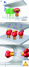
Model of the morbillivirus fusion mechanism. (A) Viral and cellular proteins involved in fusion; (B) target membrane invasion; and (C) pore formation. (A, bottom) Viral membrane (gray) with the attachment (H) protein tetrameric complex (green) and the F protein trimer. (Top) Cellular membrane (gray) with the receptor (dark blue). The H protein has a six-blade propeller structure (9, 56) and consists of two noncovalently linked dimers of covalently linked dimers (36, 37). The main receptor protein for both MV and CDV is the signaling lymphocytic activation molecule (SLAM, also called CD150) of the immunoglobulin superfamily (50, 51). The contact surface areas both on SLAM (32) and the attachment protein (45, 56) have been defined. The F protein consist of a fusion peptides (blue horizontal cylindrical structure), a short linker (light gray cylinder), HRA (yellow cylinders numbered 1 to 3), the body of the F2 and F1 subunits (large elongated red object), HRB (orange cylinders numbered 4 to 6), another short linker (a gray continuous cylinder in two monomers; an interrupted gray and blue cylinder in the cleaved monomer), and the TM segment with the cytoplasmic tail (blue thin cylindrical structure). (B) Six circularly arranged F protein trimers in the target membrane invasion conformation. (C) Trimers in their most stable six-helix bundle conformation arranged circularly around a fusion pore. Three of the six trimers have been removed for visual clarity. The inset is a coronal section of the six-helix bundle. For details, see the text.

Similar articles

Cited by

References

    1. Althoff, K., J. Mullberg, D. Aasland, N. Voltz, K. Kallen, J. Grotzinger, and S. Rose-John. 2001. Recognition sequences and structural elements contribute to shedding susceptibility of membrane proteins. Biochem. J. 353:663-672. - PMC - PubMed
    1. Baker, K. A., R. E. Dutch, R. A. Lamb, and T. S. Jardetzky. 1999. Structural basis for paramyxovirus-mediated membrane fusion. Mol. Cell 3:309-319. - PubMed
    1. Begona Ruiz-Arguello, M., L. Gonzalez-Reyes, L. J. Calder, C. Palomo, D. Martin, M. J. Saiz, B. Garcia-Barreno, J. J. Skehel, and J. A. Melero. 2002. Effect of proteolytic processing at two distinct sites on shape and aggregation of an anchorless fusion protein of human respiratory syncytial virus and fate of the intervening segment. Virology 298:317-326. - PubMed
    1. Brody, B. A., S. S. Rhee, M. A. Sommerfelt, and E. Hunter. 1992. A viral protease-mediated cleavage of the transmembrane glycoprotein of Mason-Pfizer monkey virus can be suppressed by mutations within the matrix protein. Proc. Natl. Acad. Sci. USA 89:3443-3447. - PMC - PubMed
    1. Cathomen, T., H. Y. Naim, and R. Cattaneo. 1998. Measles viruses with altered envelope protein cytoplasmic tails gain cell fusion competence. J. Virol. 72:1224-1234. - PMC - PubMed

Publication types

LinkOut - more resources