. 2004 Aug;78(15):7969-83.
doi: 10.1128/JVI.78.15.7969-7983.2004.

Intracellular processing of human herpesvirus 6 glycoproteins Q1 and Q2 into tetrameric complexes expressed on the viral envelope

Affiliations

Intracellular processing of human herpesvirus 6 glycoproteins Q1 and Q2 into tetrameric complexes expressed on the viral envelope

Pilailuk Akkapaiboon et al. J Virol. 2004 Aug.

Abstract

Human herpesvirus 6 (HHV-6) glycoproteins H and L (gH and gL, respectively) and the 80-kDa form of glycoprotein Q (gQ-80K) form a heterotrimeric complex that is found on the viral envelope and that is a viral ligand for human CD46. Besides gQ-80K, the gQ gene encodes an additional product whose mature molecular mass is 37 kDa (gQ-37K) and which is derived from a different transcript. Therefore, we designated gQ-80K as gQ1 and gQ-37K as gQ2. We show here that gQ2 also interacts with the gH-gL-gQ1 complex in HHV-6-infected cells and in virions. To examine how these components interact in HHV-6-infected cells, we performed pulse-chase studies. The results demonstrated that gQ2-34K, which is endo-beta-N-acetylglucosaminidase H sensitive and which is the precursor form of gQ2-37K, associates with gQ1-74K, which is the precursor form of gQ1-80K, within 30 min of the pulse period. After a 1-h chase, these precursor forms had associated with the gH-gL dimer. Interestingly, an anti-gH monoclonal antibody coimmunoprecipitated mainly gQ1-80K and gQ2-37K, with little gQ1-74K or gQ2-34K. These results indicate that although gQ2-34K and gQ1-74K interact in the endoplasmic reticulum, the gH-gL-gQ1-80K-gQ2-37K heterotetrameric complex arises in the post-endoplasmic reticulum compartment. The mature complex is subsequently incorporated into viral particles.


Figures

FIG. 1.
FIG. 1.
Organization of the transcription pattern of the HHV-6A U100 (gQ) gene region. (A) All exons and introns are drawn to scale. Numbering starts with the first nucleotide of the 5′ end of both transcripts gQ1 and gQ0. Numbers for splice sites apply to the last nucleotide of exon n and to the first nucleotide of exon n + 1. For special sequence motifs, the numbers apply to the first 5′ nucleotide. Thin lines represent introns; thick lines represent exons. The arrow-shaped boxes represent ORFs with ATG initiation codons and TGA termination codons. The dotted lines represent the positions of recombinant proteins (AgQ1 and AgQ2) used to produce MAbs. The single-stranded DNA probes (1R, 2R, 3R, and 4R) used for Northern blot analyses, the primers used for 5′ RACE (gQ-R, 1R, and 2R for the gQ2 transcript; gQR, 3R, and 5R for the gQ1 transcript), and the primers used for RT-PCR (gQ-F, gQ-R, gQ1-R, and gQ2-F) are indicated by small closed arrows. The sizes of the cDNA molecules and the corresponding mRNAs, identified by Northern blotting of transcripts gQ1 and gQ2, are noted on the right. The sequence of the gQ0 transcript of strain GS was from Pfeiffer et al. (31). a.a., amino acids. (B) Location of the gQ gene on the viral genome. The HHV-6 genome is shown with emphasis on the right terminal region. DRL and DRR, left and right direct repeats, respectively. (C) Amino acid (aa) sequences of the predicted products of the HHV-6A U100 genes, gQ1 and gQ2. The hydrophobic domain is underlined, and potential N-linked glycosylation sites are boxed. Numbers on the right denote amino acid residues.
FIG. 1.
FIG. 1.
Organization of the transcription pattern of the HHV-6A U100 (gQ) gene region. (A) All exons and introns are drawn to scale. Numbering starts with the first nucleotide of the 5′ end of both transcripts gQ1 and gQ0. Numbers for splice sites apply to the last nucleotide of exon n and to the first nucleotide of exon n + 1. For special sequence motifs, the numbers apply to the first 5′ nucleotide. Thin lines represent introns; thick lines represent exons. The arrow-shaped boxes represent ORFs with ATG initiation codons and TGA termination codons. The dotted lines represent the positions of recombinant proteins (AgQ1 and AgQ2) used to produce MAbs. The single-stranded DNA probes (1R, 2R, 3R, and 4R) used for Northern blot analyses, the primers used for 5′ RACE (gQ-R, 1R, and 2R for the gQ2 transcript; gQR, 3R, and 5R for the gQ1 transcript), and the primers used for RT-PCR (gQ-F, gQ-R, gQ1-R, and gQ2-F) are indicated by small closed arrows. The sizes of the cDNA molecules and the corresponding mRNAs, identified by Northern blotting of transcripts gQ1 and gQ2, are noted on the right. The sequence of the gQ0 transcript of strain GS was from Pfeiffer et al. (31). a.a., amino acids. (B) Location of the gQ gene on the viral genome. The HHV-6 genome is shown with emphasis on the right terminal region. DRL and DRR, left and right direct repeats, respectively. (C) Amino acid (aa) sequences of the predicted products of the HHV-6A U100 genes, gQ1 and gQ2. The hydrophobic domain is underlined, and potential N-linked glycosylation sites are boxed. Numbers on the right denote amino acid residues.
FIG. 2.
FIG. 2.
Transcription pattern of the U100 (gQ) region of HHV-6A strain GS with several probes. Five micrograms each of poly(A)+ RNA from mock-infected cells (Mock) and cells infected for 72 h with HHV-6A strain GS (GS) was subjected to Northern blot analysis. mRNA sizes are indicated on the right. The positions of the oligonucleotide probes used here are shown in Fig. 1A. β-Actin was used as an internal control for the detection of mRNA. 1R and 2R, antisense probes derived from the third and second exons of the gQ2 transcript, respectively, detected mRNAs of 5.6, 5.5, 3.7, 2.7, and 0.8 kb (panels 1R and 2R). In contrast, 3R and 4R, antisense probes derived from the ninth and fifth exons of the gQ1 transcript, respectively, detected mRNAs of 5.6, 5.5, 3.7, 2.9, and 2.7 kb but not the 0.8-kb mRNA (panels 3R and 4R).
FIG. 3.
FIG. 3.
Immunoblotting of recombinant gQ1- or gQ2-transfected 293T cells. 293T cells were transfected with plasmid pCAGGS-gQ1 (amplified by primers gQ-F and gQ1-R in gQ1 of Fig. 1A), plasmid pCAGGS-gQ2 (amplified by primers gQ2-F and gQ-R in gQ2 of Fig. 1A), or plasmid pCAGGS as a negative control. (A) The cells were lysed with RIPA buffer at 48 h posttransfection, resolved by SDS-PAGE with 10 or 15% polyacrylamide under reducing conditions, and electrotransferred to PVDF membranes. The blots were reacted with MAb AgQ-119 (for gQ1) (a) or MAb AgQ-182 (for gQ2) (b). (B) Lysates were digested with endo H (H) or PNGase F (F), resolved by SDS-PAGE with 15 or 7.5% polyacrylamide under reducing conditions, and electrotransferred to PVDF membranes. The blots were reacted with the MAb for gQ1 (a) or the MAb for gQ2 (b). The numbers beside the panels show molecular masses. WB, Western blotting.
FIG. 4.
FIG. 4.
Immunoblotting of HHV-6A strain GS-infected HSB-2 cells and purified virions. Lysates were digested with endo H (H) or PNGase F (F), resolved by SDS-PAGE with 10% polyacrylamide under reducing conditions, and electrotransferred to PVDF membranes. The blots were reacted with MAb AgQ-182 (for gQ2) or MAb AgQ-119 (for gQ1). The numbers beside the panels show molecular masses. (A) HSB-2 cells were infected with strain GS at an MOI of 1.0 or mock infected. The cells were lysed with RIPA buffer at 96 h postinfection. (B) Purified GS virions were lysed with RIPA buffer and digested with endo H (H) or PNGase F (F). WB, Western blotting.
FIG. 5.
FIG. 5.
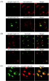
Confocal microscope analyses of strain GS-infected HSB-2 cells. Strain GS-infected HSB-2 cells were harvested 3 days postinfection and then fixed and stained for IFA. (A) MAbs AgQ-119 (for gQ1) and AgQ-182 (for gQ2) were detected with FITC-goat anti-mouse immunoglobulin G (IgG), and the anticalreticulin antibody was detected with rhodamine isothiocyanate (RITC)-swine anti-rabbit IgG. (B) MAbs AgQ-119 (for gQ1) and AgQ-182 (for gQ2) were detected with RITC-rabbit anti-mouse IgG, and lectin was detected with HPL-FITC conjugates. (C) gQ1 was detected with AgQ-119-FITC conjugates, and AgQ-182 (for gQ2) was detected with RITC-rabbit anti-mouse IgG. Specific immunofluorescence was observed with a confocal laser scanning microscope (Nikon TE300 Bio-Rad Radiance 2100 for panels A and B and Carl Zeiss LSM 510 for panel C).
FIG. 6.
FIG. 6.
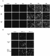
IFA of HHV-6A-infected HSB-2 cells at various times postinfection. (A) HSB-2 cells were infected with strain GS at an MOI of 0.1 or mock infected. The cells were harvested at 12, 24, 48, 72, or 96 h postinfection and stained with MAb AgQ-182 (for gQ2), 1D3 (for gH), or AgL3 (for gL). (B) HSB-2 cells were infected with strain GS at an MOI of 0.1 in the presence (+) or absence (−) of PFA. The cells were harvested at 48 h postinfection and stained with MAb AgQ-182 (for gQ2), AgQ-119 (for gQ1), or OHV-2 (for early antigen of HHV-6).
FIG. 7.
FIG. 7.
Immunoprecipitation of [35S]methionine-labeled strain GS-infected cells. (A) Mock- and HHV-6A strain GS-infected HSB-2 cells were metabolically labeled with [35S]methionine for 16 h, lysed, and immunoprecipitated (ip) with MAb AgQ-119 (for gQ1) or AgQ-182 (for gQ2). The immunoprecipitates were resolved by SDS-PAGE (4 to 12% Tris-glycine gel; Invitrogen). (B) Mock- and HHV-6A strain GS-infected HSB-2 cells were pulse-labeled with [35S]methionine for 30 min and chased for 1, 2, or 4 h. The lysates were immunoprecipitated with AgQ-119 (gQ1) or AgQ-182 (gQ2), and the immunoprecipitates were resolved by SDS-10% PAGE. Lanes: p, 30-min pulse; 1h, 1-h chase; 2h, 2-h chase; 4h, 4-h chase.
FIG. 8.
FIG. 8.
Detection of gH-gL-gQ1-gQ2 complexes in strain GS-infected HSB-2 cells. Mock- or strain GS-infected HSB-2 cell lysates were immunoprecipitated (ip) with anti-gQ1 MAb AgQ-119, anti-gQ2 MAb AgQ-182, or anti-gH MAb 1D3 and subjected to SDS-PAGE (4 to 12% Tris-glycine gel; Invitrogen) under reducing conditions. The gels were electrotransferred to PVDF membranes and probed with MAb for gQ1 (a), MAb for gQ2 (b), anti-gH monospecific serum (c), or MAb for gL (d). HC, immunoglobulin heavy chain of the immunoprecipitating antibody; LC, immunoglobulin light chain of the immunoprecipitating antibody. WB, Western blotting.
FIG. 9.
FIG. 9.
Detection of the gH-gL-gQ1-80K-gQ2-37K complex in purified virions. (A) Purified GS virion lysates were immunoprecipitated (ip) with MAb AgQ-119 (gQ1), AgQ-182 (gQ2), or 1D3 (gH) and subjected to SDS-PAGE (4 to 12% Tris-glycine gel; Invitrogen) under reducing conditions. The gels were electrotransferred to PVDF membranes and probed with MAb for gQ1 (a), MAb for gQ2 (b), anti-gH monospecific serum (c), or MAb for gL (d). (B) Purified GS virion lysates were immunoprecipitated (ip) with MAb AgQ-119 (gQ1), AgQ-182 (gQ2), or 87-Y-13 (gB) and subjected to SDS-10% PAGE under reducing conditions. The gels were electrotransferred to PVDF membranes and probed with MAb for gQ1 (a), MAb for gQ2 (b), or MAb for gB (c). HC, immunoglobulin heavy chain of the immunoprecipitating antibody; LC, immunoglobulin light chain of the immunoprecipitating antibody. WB, Western blotting.
FIG. 10.
FIG. 10.
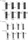
Detection of gH-gL-gQ1-gQ2 complexes in strain GS-infected cells and purified GS virions by immunoblotting under nonreducing conditions. Strain GS-infected HSB-2 cells (A) and purified GS virions (B) were solubilized and subjected to SDS-10% PAGE under reducing (a) or nonreducing (b) conditions, followed by immunoblotting. The membranes were probed with AgQ-119 (gQ1), AgQ-182 (gQ2), anti-gH monospecific serum, or AgL3 (gL). WB, Western blotting.
FIG. 11.
FIG. 11.
Model of the intracellular processing of the gH-gL-gQ1-gQ2 tetrameric complex in HHV-6-infected cells. First, gQ1-74K and gQ2-34K associate to form a heterodimeric complex in the ER. After gQ1-gQ2 dimer formation, gQ1-74K-gQ2-34K associates with gL or with gH-gL to form a gH-gL-gQ1-gQ2 complex in the post-ER compartment. Once these post-ER processing steps have been completed, the mature gH-gL-gQ1-gQ2 heterotetrameric complex is competent to traffic to compartments that will insert it into the HHV-6 envelope.

Similar articles

Cited by

References

    1. Ablashi, D. V., N. Balachandran, S. F. Josephs, C. L. Hung, G. R. Krueger, B. Kramarsky, S. Z. Salahuddin, and R. C. Gallo. 1991. Genomic polymorphism, growth properties, and immunologic variations in human herpesvirus-6 isolates. Virology 184:545-552. - PubMed
    1. Anderson, R. A., D. X. Liu, and U. A. Gompels. 1996. Definition of a human herpesvirus-6 betaherpesvirus-specific domain in glycoprotein gH that governs interaction with glycoprotein gL: substitution of human cytomegalovirus glycoproteins permits group-specific complex formation. Virology 217:517-526. - PubMed
    1. Aubin, J. T., H. Collandre, D. Candotti, D. Ingrand, C. Rouzioux, M. Burgard, S. Richard, J. M. Huraux, and H. Agut. 1991. Several groups among human herpesvirus 6 strains can be distinguished by Southern blotting and polymerase chain reaction. J. Clin. Microbiol. 29:367-372. - PMC - PubMed
    1. Campadelli-Fiume, G., S. Guerrini, X. Liu, and L. Foa-Tomasi. 1993. Monoclonal antibodies to glycoprotein B differentiate human herpesvirus 6 into two clusters, variants A and B. J. Gen. Virol. 74:2257-2262. - PubMed
    1. Chandran, B., S. Tirawatnapong, B. Pfeiffer, and D. V. Ablashi. 1992. Antigenic relationships among human herpesvirus-6 isolates. J. Med. Virol. 37:247-254. - PubMed

Publication types

LinkOut - more resources