. 2004 Aug;78(15):7990-8001.
doi: 10.1128/JVI.78.15.7990-8001.2004.

Transport of African swine fever virus from assembly sites to the plasma membrane is dependent on microtubules and conventional kinesin

Affiliations

Transport of African swine fever virus from assembly sites to the plasma membrane is dependent on microtubules and conventional kinesin

Nolwenn Jouvenet et al. J Virol. 2004 Aug.

Abstract

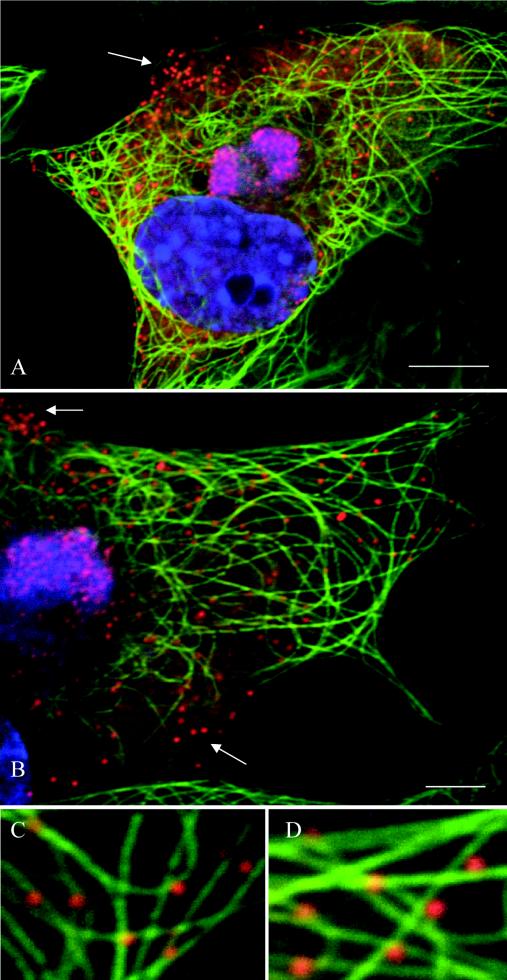
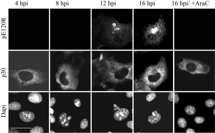
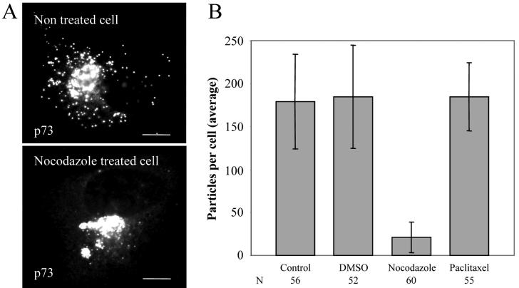
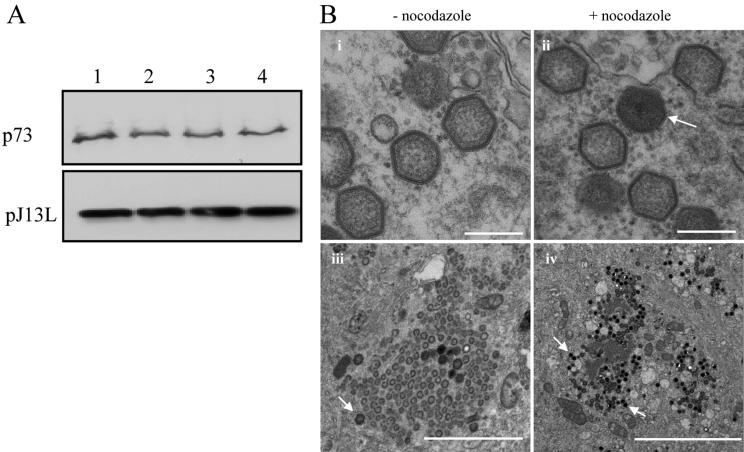
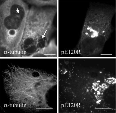
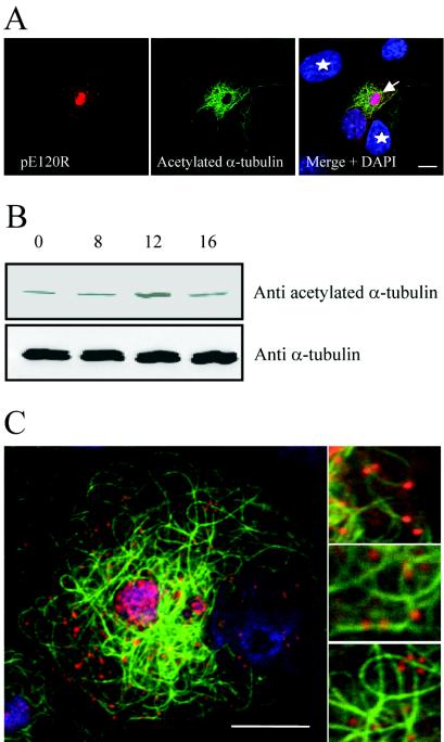
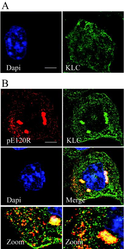
African swine fever virus (ASFV) is a large DNA virus that assembles in perinuclear viral factories located close to the microtubule organizing center. In this study, we have investigated the mechanism by which ASFV reaches the cell surface from the site of assembly. Immunofluorescence microscopy revealed that at 16 h postinfection, mature virions were aligned along microtubules. Furthermore, virus movement to the cell periphery was inhibited when microtubules were depolymerized by nocodazole. In addition, ASFV infection resulted in the increased acetylation of microtubules as well as their protection against depolymerization by nocodazole. Immunofluorescence microscopy showed that conventional kinesin was recruited to virus factories and to a large fraction of virus particles in the cytoplasm. Consistent with a role for conventional kinesin during ASFV egress to the cell periphery, overexpression of the cargo-binding domain of the kinesin light chain severely inhibited the movement of particles to the plasma membrane. Based on our observations, we propose that ASFV is recognized as cargo by conventional kinesin and uses this plus-end microtubule motor to move from perinuclear assembly sites to the plasma membrane.


Figures

FIG. 1.
FIG. 1.
ASFV particles associate with microtubules. Vero cells were infected with the Ba71v strain of ASFV and fixed at 16 hpi. Samples were incubated with a rabbit antiserum raised against the late ASFV structural protein pE120R (red) and a mouse antibody against α-tubulin (green). Viral DNA and cellular DNA were labeled with DAPI (blue). Images shown have been collected sequentially with a confocal laser scanning microscope and merged. The arrows indicate viral particles that do not align with microtubules. Panels C and D show enlarged views of parts of B. Scale bars, 8 μm (A) and 4 μm (B).
FIG. 2.
FIG. 2.
The anti-pE120R antibody recognizes newly assembled virions. Vero cells were infected with the Ba71v strain of ASFV, fixed at the indicated time, and processed for immunofluorescence. Samples were incubated with a rabbit antiserum raised against the late ASFV structural protein pE120R (top panels) and a mouse antibody against the early viral protein p30 (middle panels). Viral DNA and cellular DNA were labeled with DAPI (bottom panels). The ASFV structural protein pE120R is not detected until 12 hpi or not at all in the presence of Ara-C, an inhibitor of late gene expression. Scale bar, 20 μm.
FIG. 3.
FIG. 3.
Effect of nocodazole and paclitaxel on movement of ASFV into the cytosol. Vero cells were infected with the Ba71v strain of ASFV. Nocodazole, paclitaxel, or control (DMSO) was added at 12 hpi, and cells were incubated for a further 4 h prior to fixation and processing for immunofluorescence. Virions were located by immunofluorescence staining by using an antibody specific for the main capsid protein p73. (A) Immunofluorescence images show viral dispersion in a control cell and a cell incubated with nocodazole. Scale bar, 8 μm. (B) Cells were examined by microscopy as described above, and viral dispersion was quantified by counting individual particles located outside the factory. Results are presented as the average number of virions present in the cytoplasm per cell. Error bars indicate the standard deviations of the means. N, number of cells evaluated.
FIG. 4.
FIG. 4.
Effect of nocodazole on ASFV replication and assembly. (A) Effect of nocodazole on synthesis of viral late proteins. Vero cells were infected with ASFV, and nocodazole, paclitaxel, or control (DMSO) was added at 12 hpi. Samples were either labeled with [35S]methionine and cysteine for 30 min and immunoprecipitated 3.5 h later by using antibody specific for p73 (top) or incubated for a further 4 h and assayed for expression of pJ13L by Western blotting (bottom). Lane 1, no additions; lane 2, DMSO control; lane 3, nocodazole; lane 4, paclitaxel. (B) Effect of nocodazole on assembly of ASFV. Vero cells were infected with the Ba71v strain of ASFV. Nocodazole or control (DMSO) was added at 12 hpi, and cells were incubated for a further 4 h prior to processing for EM. Micrographs in frames i and ii show thin-section images of viruses formed in the presence (+) or absence (−) of nocodazole as indicated. Scale bar, 250 nm. Micrographs in frames iii and iv show similar images of virus assembly sites at lower magnification. The arrows indicate fully assembled virions. Scale bars, 2 μm (iii) and 5 μm (iv).
FIG. 5.
FIG. 5.
Microtubules are stabilized in cells infected with ASFV. Vero cells were infected with ASFV, and nocodazole was added at 12 hpi. Cells were incubated for a further 4 h prior to fixation and immunofluorescence microscopy analysis. Infected cells were identified by using a rabbit antiserum raised against the late ASFV structural protein pE120R, and a mouse antibody against α-tubulin was used to label microtubules. The star indicates a pE120R-negative cell, while the arrow indicates stabilized microtubules in a pE120R-positive cell. Scale bar, 8 μm. The figure compares cells with low (top panel) and high (bottom panels) numbers of stabilized microtubules.
FIG. 6.
FIG. 6.
ASFV infection induces acetylation of microtubules. (A and B) Identification of acetylated tubulin. Panel A shows Vero cells infected with the Ba71v strain of ASFV and fixed at 16 hpi. Samples were incubated with a rabbit antiserum raised against the late ASFV structural protein pE120R (red) and a mouse antibody against acetylated α-tubulin (green). Viral and cellular DNA were labeled with DAPI (blue). Stars indicate cells that are pE120R negative, and the arrow indicates an infected cell. Scale bar, 8 μm. Panel B shows Vero cells infected with the Ba71v strain of ASFV and lysed at the indicated times. Samples were analyzed by Western blotting with antibodies specific for α-tubulin or acetylated α-tubulin as indicated. (C) ASFV particles associate with acetylated microtubules. Vero cells were infected with ASFV and processed for immunofluorescence at 16 h. Samples were incubated with a rabbit antiserum raised against structural protein pE120R (red) and a mouse antibody against acetylated α-tubulin (green). Viral DNA and cellular DNA were labeled with DAPI (blue). Images shown were collected sequentially with a confocal laser scanning microscope and merged. Right panels are enlarged views of parts of the left image. Scale bar, 8 μm.
FIG. 7.
FIG. 7.
Conventional kinesin is recruited into ASFV assembly sites. (A) Distribution of kinesin light chain in noninfected cells. Noninfected Vero cells were fixed and processed for immunofluorescence by using the mouse kinesin light-chain antibody 63-90 (green). Cellular DNA was labeled with DAPI (blue). (B) Distribution of kinesin light chain in ASFV-infected cells. Vero cells were infected with the Ba71v strain of ASFV, fixed at 16 hpi, and processed for immunofluorescence. Rabbit antiserum raised against the structural protein pE120R was used to locate virus particles (red), and conventional kinesin light chain was identified by using the mouse antibody 63-90 (green). Viral DNA and cellular DNA were labeled with DAPI (blue). The bottom panels are enlarged views of the merge image. Scale bar, 8 μm.
FIG. 8.
FIG. 8.
ASFV particles bind the TPR domain of kinesin light chain. Vero cells were transfected with pEL-GFP-TPR and infected with ASFV 2 h later. Cells were processed for immunofluorescence by using antibodies specific for the late viral protein p73 (red) at the times indicated below. The subcellular localization of the TPR domain of kinesin light chain was monitored by the intrinsic fluorescence of GFP-TPR (green). (A) In the top gallery cells were fixed at 12 h and a view was chosen to show the distribution of GFP-TPR in the presence (star) or absence (arrow) of p73. Viral DNA and cellular DNA were labeled with DAPI (blue). Scale bar, 20 μm. In the bottom gallery cells were fixed at 16 h, and cells expressing low levels of GFP-TPR were selected to show the location of GFP-TPR in cells where virions were located outside the factory. Scale bar, 8 μm. (B) The same experiment was repeated, and samples were viewed at higher magnification at 12 hpi by using an antibody against pJ13L (bottom panels) to locate membranes in virus factories and an antibody specific for p73 (top panels) to identify virus particles. Scale bar, 4 μm.
FIG. 9.
FIG. 9.
Conventional kinesin is required for anterograde transport of ASFV to the cell surface. (A) Location of ASFV particles in cells expressing GFP-TPR or GFP-5AR. Vero cells were transfected with pEL-GFP-TPR or pEL-GFP-5AR as indicated. Cells were infected with ASFV 2 h later, fixed, and processed for immunofluorescence at 16 hpi. Virus particles were identified with an antibody against p73 (left panels) and the expression of GFP-TPR and GFP-5AR was monitored by the intrinsic fluorescence of GFP (right panels). Cells expressing high levels of GFP were selected. Scale bar, 20 μm. (B) Quantification of ASFV spread to the cell periphery in cells expressing GFP-TPR or GFP-5AR. Infected cells expressing pEL-GFP-TPR or pEL-GFP-5AR were prepared as described above. The numbers of virus particles present in the cytoplasm were compared with cells (control) on the same coverslip that were infected but did not express either GFP-TPR or GFP-5AR. Results are presented as the average number of virions present in the cytoplasm per cell. Error bars indicate the standard deviation of the means. N, number of cells evaluated.

Similar articles

Cited by

References

    1. Afonso, C. L., C. Alcaraz, A. Brun, M. D. Sussman, D. V. Onisk, J. M. Escribano, and D. L. Rock. 1992. Characterization of p30, a highly antigenic membrane and secreted protein of African swine fever virus. Virology 189:368-373. - PubMed
    1. Allan, V. J., H. M. Thompson, and M. A. McNiven. 2002. Motoring around the Golgi. Nat. Cell Biol. 4:E236-E242. - PubMed
    1. Alonso, C., J. Miskin, B. Hernaez, P. Fernandez-Zapatero, L. Soto, C. Canto, I. Rodriguez-Crespo, L. Dixon, and J. M. Escribano. 2001. African swine fever virus protein p54 interacts with the microtubular motor complex through direct binding to light-chain dynein. J. Virol. 75:9819-9827. - PMC - PubMed
    1. Andres, G., R. Garcia-Escudero, C. Simon-Mateo, and E. Vinuela. 1998. African swine fever virus is enveloped by a two-membraned collapsed cisterna derived from the endoplasmic reticulum. J. Virol. 72:8988-9001. - PMC - PubMed
    1. Andres, G., R. Garcia-Escudero, E. Vinuela, M. L. Salas, and J. M. Rodriguez. 2001. African swine fever virus structural protein pE120R is essential for virus transport from assembly sites to plasma membrane but not for infectivity. J. Virol. 75:6758-6768. - PMC - PubMed

Publication types

LinkOut - more resources