. 2004 Aug;78(15):8026-35.
doi: 10.1128/JVI.78.15.8026-8035.2004.

Comprehensive mutational analysis of a herpesvirus gene in the viral genome context reveals a region essential for virus replication

Affiliations

Comprehensive mutational analysis of a herpesvirus gene in the viral genome context reveals a region essential for virus replication

Anja Bubeck et al. J Virol. 2004 Aug.

Abstract

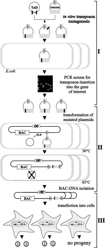
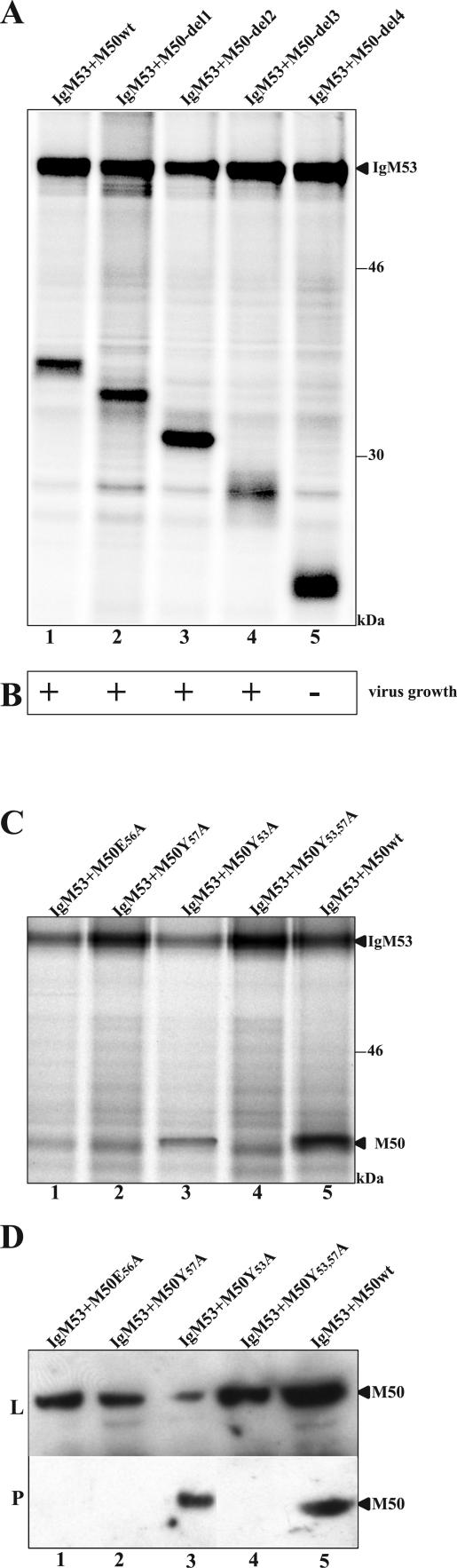
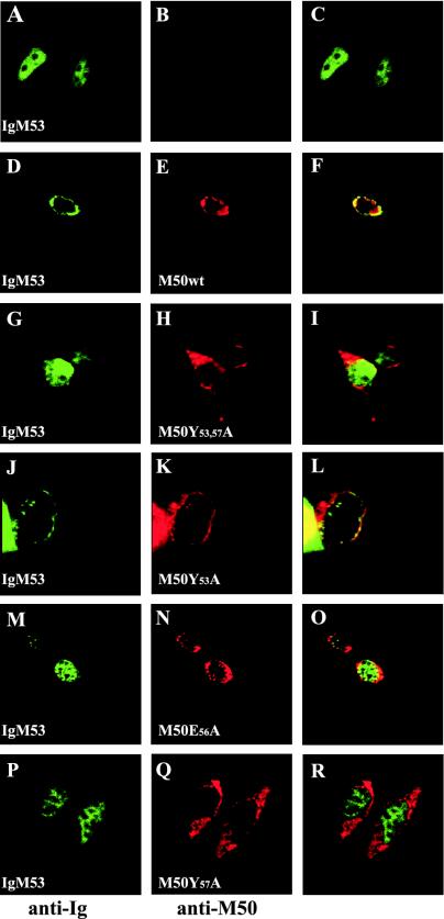
Essential viral proteins perform vital functions during morphogenesis via a complex interaction with other viral and cellular gene products. Here, we present a novel approach to comprehensive mutagenesis of essential cytomegalovirus genes and biological analysis in the 230-kbp-genome context. A random Tn7-based mutagenesis procedure at the single-gene level was combined with site-specific recombination via the FLP/FLP recognition target site system for viral genome reconstitution. We show the function of more than 100 mutants from a larger library of M50/p35, a protein involved in capsid egress from the nucleus. This protein recruits other viral proteins and cellular enzymes to the inner nuclear membrane. Our approach enabled us to rapidly discriminate between essential and nonessential regions within the coding sequence. Based on the prediction of the screen, we were able to map a site essential for viral protein-protein interaction at the amino acid level.


Figures

FIG. 1.
FIG. 1.
Strategy for random mutagenesis of an essential viral gene in the viral genome context. (I) The viral gene of interest (gray box) is subcloned into a rescue plasmid (rescue) containing one FRT site (open box with gray triangle). This plasmid is subjected to an in vitro Tn7-based random mutagenesis procedure (TnD), leading to a mutant library with 15-bp insertions (black box) into the target plasmid. This mutant library is transformed into E. coli (open boxes), and single clones are screened by PCR for insertions into the gene of interest. (II) To reinsert the gene mutants into the viral genome, the respective MCMV BAC and an FLP recombinase-expressing plasmid (FLP) are maintained in E. coli. Subsequently, the rescue plasmids are transformed into E. coli. FLP recombinase mediates site-specific recombination between the FRT sites and unifies the BAC and the rescue plasmid. Selection with chloramphenicol and Zeocin identifies BACs, with the inserted rescue plasmid carrying the mutated gene of interest. (III) Subsequently, BAC DNA is isolated and transfected into eukaryotic cells for virus reconstitution and cells are screened for viral plaques.
FIG. 2.
FIG. 2.
Ectopic rescue of the M50 deletion mutant. (A) Structure of the m152 region within pΔM50-GFP. The HindIII B fragment of BAC pΔM50-GFP contains one FRT site (pΔM50-GFP). In the HindIII H fragment of pΔM50-GFP, the M50 ORF was replaced by a kanamycin cassette (open box with X). pM50E shows this genomic region after the insertion of pOriR6Kzeo-M50 into pΔM50-GFP by site-specific recombination driven by the FLP recombinase. Arrowheads indicate the orientation of the respective ORFs. Arrows indicate HindIII restriction sites, and HindIII fragment sizes are indicated below the diagrams in kilobase pairs. DNAs of the MCMV BAC pΔm152/GFP (lane 1), pΔM50-GFP (lane2), and seven randomly picked clones of pM50E (lanes 3 to 9) were digested with HindIII and analyzed by gel electrophoresis. The sizes of the DNA bands characteristic for pM50E are indicated in kilobase pairs by arrows on the right. Marker bands are indicated on the left. (B) Growth kinetics of the viruses derived from the above-described BACs. NIH 3T3 cells were infected with viruses derived from pSMfr3 (WT-MCMV), pΔm152/GFP (m152/GFP-MCMV), or pM50E (M50E-MCMV) at a multiplicity of infection of 0.1. Supernatants of the infected cells were harvested on the indicated days, and virus titers were determined on NIH 3T3 cells by the 50% tissue culture infective dose method.
FIG. 3.
FIG. 3.
Analysis of 104 mutants with transposon insertions in the M50 ORF and screen for the functional relevance of these mutants in the viral background. (A) M50 mutants and their ability to rescue virus growth of the ΔM50 genome. Displayed is the amino acid sequence of M50. Arrowheads indicate transposon insertion sites. Open arrowheads indicate insertions leading to a stop codon; filled arrowheads represent insertions leading to the introduction of 5 aa into the M50 ORF. Dark gray arrowheads indicate mutants that rescued the ΔM50 phenotype. Light gray arrowheads indicate viruses with strongly attenuated growth. Black arrowheads indicate those mutants that were not able to rescue the ΔM50 phenotype. (B) Sequence comparison of M50/p35 homologues. The amino acid sequence of M50/p35 was aligned with those of UL50 (HCMV), R50 (rat cytomegalovirus), UL34 of HSV-1 and -2, and pseudorabies virus as well as ORF 67 of murine herpesvirus 68, herpesvirus saimiri, and human herpesvirus 8 by use of the Vector NTI AlignX program (InfoMax, Bethesda, Md.) via the BLOSUM62 similarity matrix. The depicted similarity plot was calculated using a 5-aa window size and with scores for identity and strong and weak similarities of 1.0, 0.5, and 0.2, respectively. The x axis represents the number of the amino acids of the consensus sequence. Below the diagram, a proportional representation of M50/p35 is displayed. The N-terminal region is marked in dark gray; the C-terminal region is marked in light gray. The striped box indicates the TM domain; the black box indicates the proline-rich region.
FIG. 4.
FIG. 4.
Coimmunoprecipitation of M50/p35 and M53/p38. (A) Mutants tested for the interaction between M50/p35 and M53/p38 proteins. Depicted is a schematic overview of the M50/p35 protein (see Fig. 2B). Arrows indicate the transposon insertions tested for interaction with M53/p38; long arrows represent those that lost the ability to interact with M53/p38. (B) Coimmunoprecipitation of M50/p35 and M53/p38. pCR3-M50 or pCR3-M50mut and pCR3-IgM53 were cotransfected into 293 cells. Cells were radioactively labeled and subsequently lysed. IgM53 was precipitated by protein A-Sepharose. Samples were then analyzed by SDS-PAGE. Individual mutants are characterized by their insertion sites. Arrows indicate M50/p35, M50/p35mut, and IgM53 protein bands. For a control, we used wt M50 (lane 2) and the functional M50 mutant with an insertion at aa 36 (M50-aa36).
FIG. 5.
FIG. 5.
Functional analysis of M50 mutants. (A) Coprecipitation of IgM53 and M50/p35 deletion mutants. pCR3-IgM53 and M50-del1, M50-del2, M50-del3, M50-del4, and wt M50 were cotransfected into 293 cells. Cells were radioactively labeled, and protein complexes with IgM53 were precipitated with protein A-Sepharose. Samples were analyzed by SDS-PAGE. (B) Test for functionality of the M50 deletion mutants. For functional analysis, M50-del1, M50-del2, M50-del3, and M50-del4 were reinserted into pΔM50-GFP. BAC DNA was isolated and transfected into M2-10B4 cells. Ten days postinfection, cells were screened for plaque formation. The results are shown in the row below the depicted gel. (C) Coprecipitation of IgM53 and M50/p35 mutants affecting the potential binding motif. pCR3-IgM53 plus M50E56A, M50Y57A, M50Y53A, M50Y53,57A, or wt M50 was cotransfected into 293 cells. Cells were radioactively labeled, and protein complexes were precipitated with protein A-Sepharose. Samples were analyzed by SDS-PAGE. (D) Western blot analysis after pull-down of the M50-M53 complex. pCR3-IgM53 plus wt M50 M50Y53,57A, M50Y53A, M50E56A, or M50Y57A was cotransfected into 293 cells. Samples were analyzed by SDS-PAGE and plotted on membranes. M50/p35 was detected with an M50-specific antiserum in untreated cell lysate (L) and after pull-down of the M50/IgM53 complex with protein A-Sepharose (P). M50/p35-specific bands are indicated.
FIG. 6.
FIG. 6.
Localization of M53/p38 in the presence of wtM50/p35 or mutants. NIH 3T3 cells were transiently transfected with pCR3-IgM53 alone or together with wt M50, M50Y53,57A, M50Y53A, M50E56A, or M50Y57A. Cells were stained with a fluorescein-conjugated antibody against the Ig tag to detect IgM53/p38 (green) and costained with an M50/p35-specific rabbit serum, which was detected by a Texas red-coupled secondary antibody (red). The discrepancy between the appearance of M50/p35-specific cytoplasmic staining shown here and that in reference is due to the different cell lines used.

Similar articles

Cited by

References

    1. Almazan, F., J. M. Gonzalez, Z. Penzes, A. Izeta, E. Calvo, J. Plana-Duran, and L. Enjuanes. 2000. Engineering the largest RNA virus genome as an infectious bacterial artificial chromosome. Proc. Natl. Acad. Sci. USA 97:5516-5521. - PMC - PubMed
    1. Biery, M. C., F. J. Stewart, A. E. Stellwagen, E. A. Raleigh, and N. L. Craig. 2000. A simple in vitro Tn7-based transposition system with low target site selectivity for genome and gene analysis. Nucleic Acids Res. 28:1067-1077. - PMC - PubMed
    1. Bjerke, S. L., J. M. Cowan, J. K. Kerr, A. E. Reynolds, J. D. Baines, and R. J. Roller. 2003. Effects of charged cluster mutations on the function of herpes simplex virus type 1 UL34 protein. J. Virol. 77:7601-7610. - PMC - PubMed
    1. Borst, E.-M., G. Hahn, U. H. Koszinowski, and M. Messerle. 1999. Cloning of the human cytomegalovirus (HCMV) genome as an infectious bacterial artificial chromosome in Escherichia coli: a new approach for construction of HCMV mutants. J. Virol. 73:8320-8329. - PMC - PubMed
    1. Borst, E.-M., S. Mathys, M. Wagner, W. Muranyi, and M. Messerle. 2001. Genetic evidence of an essential role for cytomegalovirus small capsid protein in viral growth. J. Virol. 75:1450-1458. - PMC - PubMed

Publication types