. 2004 Aug;78(15):8135-45.
doi: 10.1128/JVI.78.15.8135-8145.2004.

Epitope mapping of the major capsid protein of type 2 porcine circovirus (PCV2) by using chimeric PCV1 and PCV2

Affiliations

Epitope mapping of the major capsid protein of type 2 porcine circovirus (PCV2) by using chimeric PCV1 and PCV2

Porntippa Lekcharoensuk et al. J Virol. 2004 Aug.

Abstract

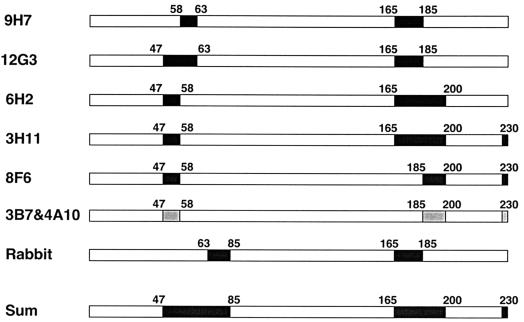
Type 2 porcine circovirus (PCV2) is associated with postweaning multisystemic wasting syndrome in pigs, whereas the genetically related type 1 PCV (PCV1) is nonpathogenic. In this study, seven monoclonal antibodies (MAbs) against PCV2-ORF2 capsid protein were generated, biologically characterized, and subsequently used to map the antigenic sites of PCV2 capsid protein by using infectious PCV DNA clones containing PCV1/PCV2-ORF2 chimeras. The PCV1/PCV2-ORF2 chimeras were constructed by serial deletions of PCV2-ORF2 and replacement with the corresponding sequences of the PCV1-ORF2. The reactivities of chimeric PCV1/PCV2 clones in transfected PK-15 cells with the seven MAbs were detected by an immunofluorescence assay (IFA). The chimera (r140) with a deletion of 47 amino acids at the N terminus of PCV2-ORF2 reacted strongly to all seven MAbs. Expanding the deletion of PCV2-ORF2 from residues 47 to 57 (r175) abolished the recognition of MAb 3B7, 3C11, 4A10, 6H2, or 8F6 to the chimera. Further deletion of PCV2-ORF2 to 62 residues disrupted the binding of this chimera to all seven MAbs. IFA reactivities with all MAbs were absent when residues 165 to 233 at the C terminus of PCV2-ORF2 was replaced with that of PCV1-ORF2. Extending the sequence of PCV2-ORF2 from residues 165 (r464) to 185 (r526), 200 (r588), or 224 (r652) restored the ability of the three chimeras to react with MAbs 3C11, 6H2, 9H7, and 12G3 but not with 8F6, 3B7, or 4A10. When the four amino acids at the C terminus of r588 were replaced with that of PCV2-ORF2, the resulting chimera (r588F) reacted with all seven MAbs. The results from this study suggest that these seven MAbs recognized at least five different but overlapping conformational epitopes within residues 47 to 63 and 165 to 200 and the last four amino acids at the C terminus of the PCV2 capsid protein.


Figures

FIG. 1.
FIG. 1.
Construction of PCV2 clone p31/31 (PCV2 strain ISU31). The ORF2 gene and the ORF1 fragment (ORF1 plus the remaining genomic sequence of PCV2) were amplified by PCR and cloned into plasmid pKSII+ (hatched line) as shown. B, X, and H are the BamHI, XhoI, and HindIII restriction sites used for cloning. The full-length PCV2 genome is excised with XhoI and recircularized with T4 DNA ligase to form a double-stranded circular DNA molecule for in vitro transfection.
FIG. 2.
FIG. 2.
Schematic diagram of PCV1/PCV2 ORF2 chimeras and their reactivities with various polyclonal and monoclonal antibodies. PCV clones p31/31 (PCV2) and p31/15 contained the complete 233-amino-acid sequences of PCV2-ORF2 (empty block) and PCV1-ORF2 (hatched block), respectively. The PCV1/PCV2 ORF2 chimeric cassettes contain serial deletions of the PCV2-ORF2 sequence joined with the remaining sequence of PCV1-ORF2 at a similar position. (A) The first set of chimeras consists of the N-terminal sequence of PCV1-ORF2 fused with the C-terminal sequence of PCV2-ORF2 at amino acid positions 14 (r39), 47 (r140), 58 (r175), 63 (r192), 85 (r256), 110 (r327), and 142 (r427), respectively. (B) For the second set of constructs, the N-terminal sequence of PCV2-ORF2 is joined with the C-terminal sequence of PCV1-ORF2 at amino acid positions 165 (r464), 185 (r526 and r526F), 200 (r588 and r588F), and 224 (r652 and r652F). The F chimeras (r526F, r588F, and r652F) differ from their corresponding ORF2 chimeras in that they contained the last four amino acid residues of PCV2-ORF2 (PLKP) rather than those of PCV1-ORF2 (-LNK) at the C terminus. (C) The third set of PCV1/PCV2 ORF2 mosaic chimeras contains the sequence of PCV2-ORF2 encompassing residues 58 to 185 (r175/526), 47 to 200 (r140/588), 47 to 200 plus the last four amino acids at the C terminus (140/588F), 47 to 224 (r652), and 47 to 224 plus the last four amino acids at C terminus (140/652F), flanked by the remaining sequence of PCV1-ORF2. (D) The last set of the chimeras contains block(s) of PCV2-ORF2 sequences at residues 47 to 62 (r140/192), 185 to 200 (r526/588), 185 to 200 plus the last four amino acids at C terminus (r526/588F), or 47 to 62 plus 185 to 200 (rbox140) flanked by PCV1-ORF2 sequences. The results of IFA reactivities between each antibody and PK-15 cells transfected with each PCV construct were indicated next to each construct. IFA reactivities of the constructs in transfected Pk-15 cells were demonstrated by PCV2 convalescent-phase swine antiserum (S), rabbit hyperimmune antiserum (R) or MAbs (9H7, 12G3, 6H2, 3C11, 8F6, 3B7, and 4A10). +, a strong reactivity; +w, a weak reactivity; +I, an intermediate or moderate reactivity; −, no reactivity.
FIG. 3.
FIG. 3.
IFA reactivity with polyclonal antibodies or a representative MAb 3B7 in PK-15 cells transfected with PCV clone p31/31 or p31/15. The upper panel indicates reactivities of PCV2 convalescent-phase swine antiserum (A), rabbit hyperimmune antiserum (B), or MAb 3B7 (C) with cells transfected with PCV2 clone p31/31. The lower panel showed reactivities between clone p31/15 and the swine antiserum (D) and no reactivity between clone p31/15 and the rabbit hyperimmune serum (E) or MAb 3B7 (F).
FIG. 4.
FIG. 4.
Comparison between deduced amino acid sequences of PCV1-ORF2 (derived from PK-15 cells) and PCV2-ORF2 (from strain ISU31 used in the study). Both PCV1-ORF2 and PCV2-ORF2 contained 233 amino acid residues. Dots (.) represent an identical amino acid in both strains. Dashes (-) represent deletions. The recombination position of each chimera is indicated by an underscore (_) either preceded or followed by the name of each chimera. The chimeras are named according to the nucleotide position at the joining junction.
FIG. 5.
FIG. 5.
Positive reactivity between each MAb and each PCV chimera. MAb 9H7 gives a strong positive signal with chimeras r140 (A), r175 (B), r526 (C), and r175/526 (D). MAb 12G3 reacts strongly with chimeras r140 (E) and r526 (F) but only weakly with r175 (G) and r175/526 (H). MAb 6H2 shows strong IFA reactivity with chimeras r140 (I), r588 (J), and r140/588 (K). MAb 8F6 also reacts strongly with chimeras r140 (L) and r588F (M). MAb 3C11 reacts strongly with chimera r140 (N), moderately with chimeras r588F (O) and r140/588F (P), and weakly with chimeras r588 (Q), r652 (R), and r140/588 (S). MAbs 4A10 and 3B7 have a similar IFA staining pattern: a strong reactivity of MAb 3B7 with chimera r140 (T) but weak reactivity with chimera r588F (U); a strong reactivity between MAb 4A10 and chimera r140 (V) but a weak reactivity with chimera r588F (W).
FIG. 6.
FIG. 6.
Schematic diagram of immunoreactive epitopes on the PCV2 capsid protein responsible for binding of polyclonal and monoclonal antibodies. Each bar represents a full-length PCV2 capsid protein. Filled boxes within bars are the regions important for epitope recognitions of MAbs. All seven MAbs recognized different but overlapping conformational epitopes comprised of amino acid residues that reside in both N- and C-terminal regions of the PCV2 capsid protein. The regions recognized by seven MAbs are as follows: amino acids 58 to 62 and 165 to 185 for MAb 9H7; amino acids 47 to 62 and 165 to 185 for MAb 12G3; amino acids 47 to 57 and 165 to 200 for MAb 6H2; amino acids 47 to 57, 165 to 200, and 230 to 233 for MAb 3C11; and amino acids 47 to 57, 185 to 200, and 230 to 233 for MAbs 8F6, 3B7, and 4A10. Reactivities between the anti-PCV2 rabbit hyperimmune antiserum and PCV chimeras reveal an additional immunoreactive region within amino acids 63 to 84 on the capsid protein. The collective reactivities (Sum) of seven MAbs and the rabbit hyperimmune antiserum with the PCV chimeras indicates that the immunodominant epitopes encompassed amino acids 47 to 84, 165 to 200, and the last four amino acids at the C terminus of the PCV2 capsid protein.

Similar articles

Cited by

References

    1. Agbandje-McKenna, M., A. L. Llamas-Saiz, F. Wang, P. Tattersall, and M. G. Rossmann. 1998. Functional implications of the structure of murine parvovirus, minute virus of mice. Structure 6:1369-1381. - PubMed
    1. Allan, G. M., F. McNeilly, J. P. Cassidy, G. A. C. Reilly, B. A. Dair, W. A. Ellis, and M. S. McNulty. 1995. Pathogenesis of porcine circovirus; experimental infections of colostrum deprived piglets and examination of pig foetal material. Vet. Microbiol. 44:49-64. - PubMed
    1. Allan, G. M., F. McNeilly, S. Kennedy, B. Daft, E. G. Clarke, J. A. Ellis, D. M. Haines, B. M. Meehan, and B. M. Adair. 1998. Isolation of porcine circovirus-like viruses from pigs with a wasting disease in the USA and Europe. J. Vet. Diagn. Investig. 10:3-10. - PubMed
    1. Allan, G. M., E. McNeilly, S. Kennedy, B. Meehan, D. Moffett, F. Malone, J. Ellis, and S. Krakowka. 2000. PCV-2-associated PDNS in Northern Ireland in 1990. Vet. Rec. 146:711-712. - PubMed
    1. Cheung, A. K. 2003. The essential and nonessential transcription units for viral protein synthesis and DNA replication of porcine circovirus type 2. Virology 313:452-459. - PubMed

Publication types

LinkOut - more resources