. 2004 Aug;78(15):8146-58.
doi: 10.1128/JVI.78.15.8146-8158.2004.

Role of nucleolin in human parainfluenza virus type 3 infection of human lung epithelial cells

Affiliations

Role of nucleolin in human parainfluenza virus type 3 infection of human lung epithelial cells

Santanu Bose et al. J Virol. 2004 Aug.

Abstract

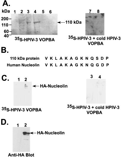
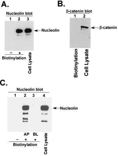
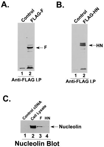
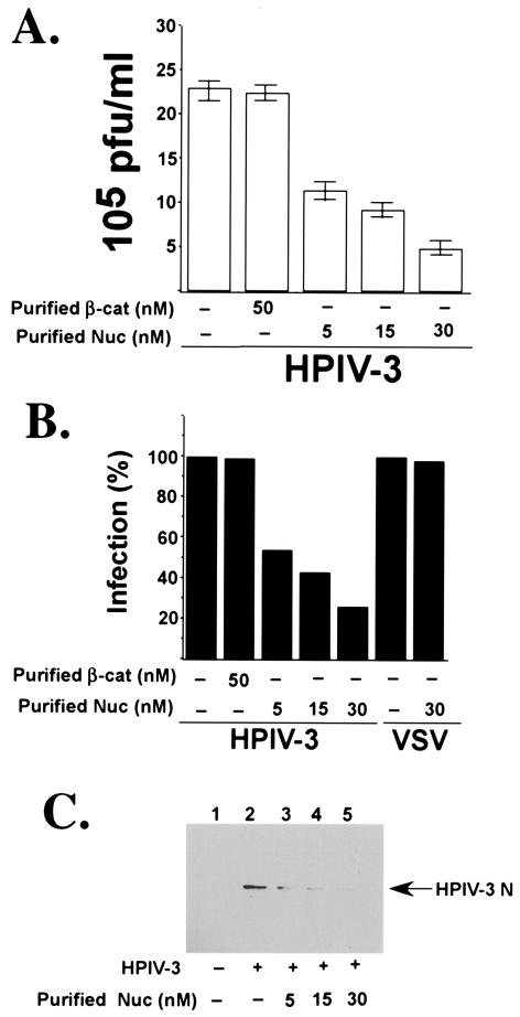
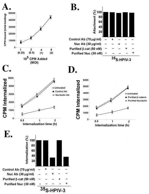
Human parainfluenza virus type 3 (HPIV-3) is an airborne pathogen that infects human lung epithelial cells from the apical (luminal) plasma membrane domain. In the present study, we have identified cell surface-expressed nucleolin as a cellular cofactor required for the efficient cellular entry of HPIV-3 into human lung epithelial A549 cells. Nucleolin was enriched on the apical cell surface domain of A549 cells, and HPIV-3 interacted with nucleolin during entry. The importance of nucleolin during HPIV-3 replication was borne out by the observation that HPIV-3 replication was significantly inhibited following (i). pretreatment of cells with antinucleolin antibodies and (ii). preincubation of HPIV-3 with purified nucleolin prior to its addition to the cells. Moreover, HPIV-3 cellular internalization and attachment assays performed in the presence of antinucleolin antibodies and purified nucleolin revealed the requirement of nucleolin during HPIV-3 internalization but not during attachment. Thus, these results suggest that nucleolin expressed on the surfaces of human lung epithelial A549 cells plays an important role during HPIV-3 cellular entry.


Figures

FIG. 1.
FIG. 1.
Identification of nucleolin as an HPIV-3 envelope binding protein. (A) VOPBA of A549 protein fractions eluted from the anion exchange column with 35S-HPIV-3 in the absence (lanes 1 to 6) and presence (lanes 7 and 8) of excess nonradioactive (cold) HPIV-3. The 35S-HPIV-3 interacting 110-kDa protein band is marked. (B) Comparison of the amino-terminal primary sequence of human nucleolin with the sequence of the 110-kDa protein. (C) 35S-HPIV-3 VOPBA in the absence (lanes 1 and 2) and presence (lanes 3 and 4) of excess nonradioactive (cold) HPIV-3 was performed by using anti-HA immunoprecipitated cell lysates obtained following transfection with HA-nucleolin (lanes 2 and 4) or an empty vector (lanes 1 and 3). (D) Western blot analysis of cell lysates (10 μg of protein) obtained from cells transfected with HA-nucleolin (lane 2) or an empty vector (lane 1) with anti-HA antibody.
FIG. 2.
FIG. 2.
Cell surface expression of nucleolin. (A) Nonbiotinylated (lane 1) and biotinylated (lane 2) A549 cell lysates (100 μg of protein) were subjected to precipitation with avidin-agarose and Western blot analysis with antinucleolin antibody. A549 cell lysates (lane 3) (4 μg of protein) served as a control. (B) Biotinylated A549 cell lysates (lane 1) (200 μg protein) were subjected to precipitation with avidin-agarose and Western blot analysis with anti-β-catenin antibody. A549 cell lysates (lane 2) (5 μg of protein) served as a control. (C) Nonbiotinylated (lane 1) and biotinylated cell lysates (250 μg protein) obtained following biotinylation from either the apical (AP) (lane 2) or the basolateral (BL) (lane 3) side of a filter-grown polarized monolayer of A549 cells were subjected to precipitation with avidin-agarose and Western blot analysis with antinucleolin antibody. Cell lysates (lane 4) (12 μg of protein) from polarized A549 cells served as a control.
FIG. 3.
FIG. 3.
Interaction of nucleolin with biotinylated HPIV-3. (A) Lysates obtained from A549 cells incubated with biotinylated HPIV-3 (Biotin-HPIV-3) (2 MOI) at 37°C were immunoprecipitated with either β-catenin (lane 1) or nucleolin (lane 2) antibodies. The bound proteins were then subjected to SDS-7.5% PAGE and blotting with avidin-HRP. Biotinylated HPIV-3 (lane 3) served as a control to demonstrate the biotinylated envelope proteins F and HN. (B) Lysates obtained from A549 cells incubated in the absence (lane 1) or presence (lane 2) of biotinylated HPIV-3 (Biotin-HPIV-3) (2 MOI) at 37°C were precipitated with avidin-agarose. The bound proteins were then subjected to Western blotting with antinucleolin antibody. A549 cell lysate (lane 3) served as a control. (C) Lysates obtained from A549 cells incubated in the absence (lane 1) or presence (lane 2) of biotinylated VSV (Biotin-VSV) (2 MOI) at 37°C were precipitated with avidin-agarose. The bound proteins were then subjected to Western blotting with antinucleolin antibody. A549 cell lysate (lane 3) served as a control. (D) Lysates obtained from A549 cells incubated in the absence (lane 1) or presence (lane 2) of biotinylated HPIV-3 (Biotin-HPIV-3) (2 MOI) at 4°C were precipitated with avidin-agarose. The bound proteins were then subjected to Western blotting with antinucleolin antibody. A549 cell lysate (lane 3) served as a control.
FIG. 4.
FIG. 4.
Interaction of nucleolin with HPIV-3 F and HN proteins. (A) A549 cells transfected with either an empty vector (lane 1) or HPIV-3 FLAG-F cDNA (lane 2) were pulse labeled with [35S]methionine, and the radioactive lysate was immunoprecipitated with anti-FLAG antibody prior to SDS-7.5% PAGE and fluorography. (B) A549 cells transfected with either an empty vector (lane 1) or HPIV-3 FLAG-HN cDNA (lane 2) were pulse labeled with [35S]methionine, and the radioactive lysate was immunoprecipitated with anti-FLAG antibody prior to SDS-7.5% PAGE and fluorography. (C) Lysates (100 μg of protein) obtained from A549 cells transfected with either an empty vector (lane 1), HPIV-3 FLAG-F (lane 3) or HPIV-3 FLAG-HN (lane 4) cDNA were immunoprecipitated with anti-FLAG antibody. The proteins bound to the washed anti-FLAG-agarose beads were subjected to Western blot analysis with antinucleolin antibody. An A549 cell lysate (lane 2) (20 μg of protein) served as a control.
FIG. 5.
FIG. 5.
Effect of nucleolin antibodies (Ab) on virus replication. (A) Culture supernatants collected from A549 cells mock infected or infected with HPIV-3 (0.2 MOI) in the absence or presence of nucleolin (Nuc) polyclonal (Poly) and monoclonal (Mono) antibodies or β-catenin polyclonal antibody (control) were added to CV-1 cells for a plaque assay. The plaque assay results, reflecting the viral titers, are expressed in PFU per milliliter. Each value represents the mean ± standard deviation for three determinations. (B) The average plaque assay values (in PFU per milliliter) from panel A were used to show the percentages of inhibition of infection in the presence of nucleolin antibodies. The percentage of infection, reflecting the percentage of virus release, was calculated as a ratio of the PFU-per-milliliter value obtained for cells infected with HPIV-3 in the presence of the antibodies to the value obtained for cells infected with HPIV-3 in the absence of the antibodies. The 100% level of infection represents the value (20 × 105 PFU/ml) obtained for untreated cells. (C) A549 cell lysates (10 μg of protein) obtained from mock-infected (lane 1) and HPIV-3-infected cells (36 h postinfection) in the absence (lane 2) or in the presence of β-catenin (control) (lane 3) or nucleolin (Nuc) (lane 4) antibodies were subjected to Western blot analysis with HPIV-3 anti-RNP antibody. (D) Culture supernatants collected from A549 cells mock infected or infected with VSV (0.2 MOI) in the absence or presence of nucleolin (Nuc) polyclonal (Poly) and monoclonal (Mono) antibodies were added to L929 cells for a plaque assay. The percentage of infection, reflecting the percentage of virus release, was calculated as a ratio of the PFU-per-milliliter value obtained for cells infected with VSV in the presence of the antibodies to the value obtained for cells infected with VSV in the absence of the antibodies. The 100% infection level represents the value (in PFU per milliliter) obtained for untreated cells. (E) A549 cell lysates (10 μg of protein) obtained from mock-infected (lane 1) and VSV-infected cells (36 h postinfection) in the absence (lane 2) or in the presence of β-catenin (control) (lane 3) or nucleolin (Nuc) (lane 4) antibodies were subjected to Western blot analysis with VSV anti-P antibody.
FIG. 6.
FIG. 6.
Effect of purified nucleolin on virus replication. (A) Culture supernatants collected from A549 cells mock infected or infected with HPIV-3 (0.2 MOI) preincubated with either purified nucleolin (Nuc) or β-catenin (β-cat) were added to CV-1 cells for a plaque assay. The plaque assay results, reflecting the viral titers, are expressed in PFU per milliliter. Each value represents the mean ± standard deviation for three determinations. (B) The average plaque assay values (in PFU per milliliter) from panel A were used to show the percentages of inhibition of infection of HPIV-3 in the presence of purified nucleolin. The percentage of infection, reflecting the percentage of virus release, was calculated as a ratio of the PFU-per-milliliter value obtained for cells infected with HPIV-3 in the presence of purified proteins to the value obtained for cells infected with HPIV-3 in the absence of purified proteins. The 100% level of infection represents the value (23 × 105 PFU/ml) obtained for untreated cells. Similarly, culture supernatants collected from A549 cells mock infected or infected with VSV (0.2 MOI) in the absence or presence of purified nucleolin were added to L929 cells for a plaque assay. The percentage of infection, reflecting the percentage of virus release, was calculated as a ratio of the PFU-per-milliliter value obtained for cells infected with VSV in the presence of the purified protein to the value obtained for cells infected with VSV in the absence of the purified protein. The 100% level of infection represents the value (in PFU per milliliter) obtained from un-treated cells. (C) A549 cell lysates (5 μg of protein) obtained from mock-infected (lane 1) and HPIV-3-infected (36 h postinfection) cells following preincubation in the absence (lane 2) or presence of 5 nM (lane 3), 15 nM (lane 4), and 30 nM (lane 5) purified nucleolin (Nuc) were subjected to Western blot analysis with HPIV-3 anti-RNP antibody.
FIG. 7.
FIG. 7.
Effect of nucleolin antibodies (Ab) and purified nucleolin (Nuc) on cellular attachment and internalization of HPIV-3. (A) The kinetics of 35S-HPIV-3 attachment to A549 cells was examined by adding different amounts of virus (0.25 to 2 MOI, or 1 × 10⁵ to 8 × 10⁵ cpm) to chilled A549 cells. Following attachment at 4°C for 2 h, the cells were washed extensively and the cell-associated radioactivity (in counts per minute) representing the attached virus was measured by counting the cell lysate with a liquid scintillation counter. Each value represents the mean ± standard deviation for three determinations. (B) Attachment of 35S-HPIV-3 (1 MOI, or 4 × 10⁵ cpm) to chilled A549 cells pretreated with antibodies or the virus preincubated with the purified proteins was determined following the attachment of the virus at 4°C for 2 h. Following adsorption, the cells were washed extensively and the cell-associated radioactivity (in counts per minute) representing the attached virus was measured by counting the cell lysate with a liquid scintillation counter. The percentage of attachment was calculated as a ratio of the amount of radioactivity present in cells incubated with 35S-HPIV-3 in the presence of the antibodies or purified proteins to the amount of radioactivity present in cells incubated with 35S-HPIV-3 alone. (C) Internalization of 35S-HPIV-3 (1 MOI, or 4 × 10⁵ cpm) into A549 cells pretreated with antibodies was determined following incubation of attached (2 h, 4°C) virus at 37°C for 0.5, 1, and 2 h. The cell-associated radioactivities (in counts per minute) representing the internalized virus at different time points were measured by counting the cell pellet with a liquid scintillation counter. Each value represents the mean ± standard deviation for three determinations. (D) Internalization of 35S-HPIV-3 (1 MOI, or 4 × 10⁵ cpm) into A549 cells following preincubation of the virus with the purified proteins was determined following incubation of attached (2 h, 4°C) virus at 37°C for 0.5, 1, and 2 h. The cell-associated radioactivities (in counts per minute) representing the internalized virus at different time points were measured by counting the cell pellet with a liquid scintillation counter. Each value represents the mean ± standard deviation for three determinations. (E) The average internalization values (counts per minute internalized) at 2 h postinternalization at 37°C from panels C and D were used to show the percentages of inhibition of internalization in the presence of nucleolin antibody and purified nucleolin. The percentage of internalization was calculated as a ratio of the amount of radioactivity present in cells infected with 35S-HPIV-3 in the presence of the antibodies or purified proteins to the amount of radioactivity present in cells infected with 35S-HPIV-3 alone. β-cat, β-catenin.

References

    1. Ah-Type, C., K. Schwartz, E. Huberman, E. Carlin, and A. Moscona. 1999. Virus-receptor interactions of human parainfluenza viruses type 1, 2, and 3. Microb. Pathog. 27:329-336. - PubMed
    1. Allain, F. H., P. Bouvet, T. Dieckmann, and J. Feigon. 2000. Molecular basis of sequence recognition of pre-ribosomal RNA by nucleolin. EMBO J. 19:6870-6881. - PMC - PubMed
    1. Bacharach, E., J. Gonsky, K. Alin, M. Orlova, and S. P. Goff. 2000. The carboxy-terminal fragment of nucleolin interacts with the nucleocapsid domain of retroviral gag proteins and inhibits virion assembly. J. Virol. 74:11027-11039. - PMC - PubMed
    1. Barton, E., C. Forrest, J. Connolly, J. Chappel, Y. Liu, F. Schnell, A. Nusrat, C. Parkos, and T. Dermody. 2001. Junction adhesion molecule is a receptor for reovirus. Cell 104:441-451. - PubMed
    1. Benito-Salas, J. S., and R. M. del Angel. 1997. Identification of two surface proteins from C6/36 cells that bind dengue type 4 virus. J. Virol. 71:7246-7252. - PMC - PubMed

Publication types