. 2004 Aug;78(15):8172-82.
doi: 10.1128/JVI.78.15.8172-8182.2004.

Calicivirus 3C-like proteinase inhibits cellular translation by cleavage of poly(A)-binding protein

Affiliations

Calicivirus 3C-like proteinase inhibits cellular translation by cleavage of poly(A)-binding protein

Muge Kuyumcu-Martinez et al. J Virol. 2004 Aug.

Abstract

Caliciviruses are single-stranded RNA viruses that cause a wide range of diseases in both humans and animals, but little is known about the regulation of cellular translation during infection. We used two distinct calicivirus strains, MD145-12 (genus Norovirus) and feline calicivirus (FCV) (genus Vesivirus), to investigate potential strategies used by the caliciviruses to inhibit cellular translation. Recombinant 3C-like proteinases (r3CL(pro)) from norovirus and FCV were found to cleave poly(A)-binding protein (PABP) in the absence of other viral proteins. The norovirus r3CL(pro) PABP cleavage products were indistinguishable from those generated by poliovirus (PV) 3C(pro) cleavage, while the FCV r3CL(pro) products differed due to cleavage at an alternate cleavage site 24 amino acids downstream of one of the PV 3C(pro) cleavage sites. All cleavages by calicivirus or PV proteases separated the C-terminal domain of PABP that binds translation factors eIF4B and eRF3 from the N-terminal RNA-binding domain of PABP. The effect of PABP cleavage by the norovirus r3CL(pro) was analyzed in HeLa cell translation extracts, and the presence of r3CL(pro) inhibited translation of both endogenous and exogenous mRNAs. Translation inhibition was poly(A) dependent, and replenishment of the extracts with PABP restored translation. Analysis of FCV-infected feline kidney cells showed that the levels of de novo cellular protein synthesis decreased over time as virus-specific proteins accumulated, and cleavage of PABP occurred in virus-infected cells. Our data indicate that the calicivirus 3CL(pro), like PV 3C(pro), mediates the cleavage of PABP as part of its strategy to inhibit cellular translation. PABP cleavage may be a common mechanism among certain virus families to manipulate cellular translation.


Figures

FIG. 1.
FIG. 1.
Comparison of the cleavage activity of NoV r3CLpro with that of PV 3Cpro and 2Apro on the cellular protein substrates, PABP and eIF4G. (A) The structural organization of human PABP and the previously mapped cleavage sites recognized by the PV 3C and 2A proteinases (32, 41). (B and C) NoV r3CLpro cleaves PABP. HeLa S3 cells were fractionated into cytoplasmic (S10) (B) and ribosome-associated fractions (C). In vitro cleavage reactions were assembled in these fractions and proteins were analyzed by SDS-10% PAGE followed by immunoblotting with PABP-specific antibodies. Numbers on the right show the migration of molecular weight markers (in thousands). HeLa S10 lysates (B) were incubated for 3.5 h at 37°C with the following: lane 1, mock incubation control (indicated by C); lane 2, heat-inactivated NoV proteinase, 3CL-In; lane 3, NoV r3CLpro, 80 ng/μl; lane 4, PV r3Cpro, 40 ng/μl; and lane 5, PV r3Cpro, 80 ng/μl. The HeLa ribosomal fraction (C) was incubated for 3.5 h at 37°C with the following: lane 1, mock control (indicated by C); lane 2, PV r3Cpro, 40 ng/μl; lane 3, PV r3Cpro, 80 ng/μl; and lane 4, NoV r3CLpro, 80 ng/μl. (D) NoV 3CLpro does not cleave eIF4G. Prior to immunoblot analysis with eIF4G antibody, HeLa S10 lysates were incubated for 3.5 h at 37°C with the following: lane 1, mock control (indicated by C); lane 2, PV r3Cpro, 40 ng/μl; lane 3, PV r3Cpro, 80 ng/μl; lane 4, NoV r3CLpro, 40 ng/μl; lane 5, NoV r3CLpro, 80 ng/μl; lane 6, CVB3 r2Apro, 40 ng/μl; and lane 7, CVB3 r2Apro, 80 ng/μl. The 115-kDa N-terminal cleavage fragment of eIF4G is indicated as cpN.
FIG. 2.
FIG. 2.
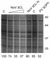
Inhibition of cellular mRNA translation by NoV r3CLpro. Autoradiogram of a 10% (wt/vol) polyacrylamide SDS-PAGE gel showing [35S]methionine-cysteine incorporation into newly synthesized proteins in HeLa translation extracts that were treated with buffer only (lanes marked C), increasing concentrations of NoV r3L Cpro at 20, 40, or 80 ng/μl (lanes 2 to 4, respectively), inactivated NoV r3CL (lane 5), or PV r3CLpro at 60 ng/μl (lane 7). Migration of molecular mass standards (in kilodaltons) is shown on the right. Relative translation intensity compared to control was determined by densitometry (percentage of control) and is shown below each lane.
FIG. 3.
FIG. 3.
NoV 3CLpro specifically inhibits poly(A)-dependent translation, and translation is restored by the addition of PABP. (A) Effect of NoV 3CLpro on translation efficiency of polyadenylated mRNA. Graphic representation of luciferase activity generated (relative light units [RLU] set to 100% of control reactions) using capped and polyadenylated luciferase RNA (left panel) or nonpolyadenylated luciferase RNA (right panel) in HeLa translation extracts incubated with increasing concentrations of NoV r3CLpro. Data represent the means ± standard deviations of four individual experiments. (B) Addition of exogenous PABP restores translation efficiency of polyadenylated mRNA in the presence of NoV 3CLpro. Comparison of polyadenylated (left panel) or nonpolyadenylated (right panel) luciferase RNA translation in HeLa translation extracts pretreated with buffer or 10 ng of NoV r3CLpro/μl and supplemented with His-PABP. Black bars represent the percent translation (light units relative to buffer control) in 3CLpro-treated lysates. Gray bars indicate luciferase translation levels after addition of His-PABP. Data represent the means ± standard deviations of three individual experiments.
FIG. 4.
FIG. 4.
Comparison of proteins produced during FCV infection with those produced in mock-infected cells over time. (A) Analysis of proteins by SDS-PAGE and autoradiography. Feline kidney cells (4 × 10⁶ cells) were either infected with FCV at an MOI of 10 or mock infected. After 3 h and then at subsequent hourly time points, medium was removed and the cells were pulse-radiolabeled for 1.5 h. The cells were then collected, washed, and boiled in SDS sample buffer prior to analysis by SDS-PAGE in a 12% polyacrylamide gel and autoradiography. Viral proteins ProPol, capsid, p39, and p32 are indicated. The asterisks illustrate two cell-associated proteins that decrease in intensity over time during FCV infection. (B) Phosphorimager quantitation of signals associated with selected cellular and viral proteins. Bands 1 and 2 correspond to the cellular proteins indicated with an asterisk in panel A, with band 1 corresponding to the slower migrating protein. The signals generated by the cellular proteins were compared to that of the 60-kDa mature FCV capsid protein. RLU, relative light units.
FIG. 5.
FIG. 5.
Analysis of PABP cleavage in FCV-infected cells and in vitro with the FCV 3CL proteinase (rProPol). (A) Comparison of PABP cleavage between PV-infected HeLa cells and FCV-infected CRFK cells. CRFK cells were either mock or FCV infected and harvested after 6 hpi. HeLa cells were mock or PV infected (MOI of 3) and harvested 6 hpi. An aliquot of FCV-infected lysates (lane 4) was compared to a PV-infected HeLa cell lysate (lane 2) by SDS-10% PAGE followed by immunoblotting with human PABP-specific antibodies. Numbers on the right show the migration of molecular weight markers (in thousands). Previously identified PABP cleavage fragments are indicated (3C, 61-kDa N-terminal cleavage fragment of PABP; 3Calt, 46-kDa N-terminal cleavage fragment of PABP; 2A, 57-kDa N-terminal cleavage fragment of PABP generated by 2Apro; FCV 3CLpro cp, FCV-induced cleavage fragment of PABP). (B) Comparison of PV 3Cpro and FCV 3CLpro in an in vitro cleavage assay of rPABP. RNA transcripts encoding PABP were translated in a reaction mixture containing 35S-labeled methionine. Translation mixtures were then incubated with 10 ng of PV 3Cpro/μl (lane 2) or FCV rProPol (lane 3) at 30°C. The translation mixture incubated without proteinase was used as negative control (indicated by C, lane 1). Marker proteins corresponding to three N-terminal fragments of PABP were produced by translation of three PABP RNAs bearing individual stop codons at positions 537 (PABP537), 487 (PABP487), or 413 (PABP413) are included as a control (lane 4). These correspond to N-terminal PABP cleavage fragments released by cleavage at the 3C, 2A, or 3Calt sites, respectively. (C) Mapping of the FCV 3CLpro cleavage site on PABP by site-directed mutagenesis of the Q413/T414, Q437/T438, and Q537/G538 dipeptides. For each construct indicated above the gel (PABP, Q413A, Q437A, and Q537A), 4 μl of a 25-μl reaction mixture was either incubated with 6 μl of PBS, pH 7.4, (odd-numbered lanes and minus signs) or 6 μl of PBS containing 5 μg of FCV 3CL (rProPol) (even-numbered lanes and plus signs). Reaction mixtures were incubated overnight at 30°C prior to separation in a 12% Tris-Gly polyacrylamide gel and autoradiography. To assess calicivirus protease activity, a MD145-12 NoV ProPol substrate (76 kDa) was translated and incubated alone or with proteases similarly (lanes 9 to 11). Molecular weight markers (in thousands) are indicated on the left. (D) FCV 3CL inhibits cellular mRNA translation in vitro. HeLa lysates were treated with buffer (indicated by C) or increasing amounts of FCV 3CL (10, 20, and 40 ng/μl) as described in Materials and Methods. An autoradiogram of SDS-PAGE is shown and percent translation quantified by densitometry is shown below the lanes. Migration of molecular mass standards (in kDa) is shown on the right.
FIG. 6.
FIG. 6.
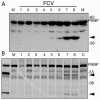
Kinetic analysis of PABP cleavage in FCV-infected cells. CRFK cells were infected with FCV as described previously, and cells were isolated at hourly time points (indicated above lanes) postinfection for analysis. (A) Immunoblot analysis of feline PABP over time in FCV-infected cells detected with antibodies raised against human PABP. An analysis of mock-infected cells harvested after 8 h incubation is shown (M). (B) Analysis of FCV-infected cell lysates in the in vitro assay for human PABP cleavage. Aliquots of infected cell extracts from time points 1 to 8 hpi were mixed with radiolabeled human PABP and incubated for 3.5 h at 30°C. Controls include radiolabeled PABP incubated with recombinant FCV ProPol enzyme, mock-infected cell extract (M), and buffer alone (C). Autoradiogram of SDS-polyacrylamide gel is shown. Arrowheads indicate PABP cleavage products. Migration of molecular weight markers is indicated on the right. The human PABP-specific antibody used in the immunoblot (A) is peptide-specific (sequence located in RRM4) and recognizes only the amino-terminal cleavage product of PABP.

Similar articles

Cited by

References

    1. Allaire, M., M. M. Chernaia, B. A. Malcolm, and M. N. G. James. 1994. Picornaviral 3C cysteine proteinases have a fold similar to chymotrypsin-like serine proteinases. Nature 369:72-76. - PubMed
    1. Al-Molawi, N., V. A. Beardmore, M. J. Carter, G. E. Kass, and L. O. Roberts. 2003. Caspase-mediated cleavage of the feline calicivirus capsid protein. J. Gen. Virol. 84:1237-1244. - PubMed
    1. Asselbergs, F., W. Peters, W. van Venrooij, and H. Bloemendal. 1978. Diminished sensitivity of re-initiation of translation to inhibition by cap analogues in reticulocyte lysates. Eur. J. Biochem. 88:483-488. - PubMed
    1. Bazan, J. F., and R. J. Fletterick. 1988. Viral cysteine proteases are homologous to the trypsin-like family of serine proteases: structural and functional implications. Proc. Natl. Acad. Sci. USA 85:7872-7876. - PMC - PubMed
    1. Belliot, G., S. V. Sosnovtsev, T. Mitra, C. Hammer, M. Garfield, and K. Y. Green. 2003. In vitro proteolytic processing of the MD145 norovirus ORF1 nonstructural polyprotein yields stable precursors and products similar to those detected in calicivirus-infected cells. J. Virol. 77:10957-10974. - PMC - PubMed

Publication types

MeSH terms

LinkOut - more resources