

Disparate regions of envelope protein regulate syncytium formation versus spongiform encephalopathy in neurological disease induced by murine leukemia virus TR
- PMID: 15254211
- PMCID: PMC446142
- DOI: 10.1128/JVI.78.15.8392-8399.2004
Disparate regions of envelope protein regulate syncytium formation versus spongiform encephalopathy in neurological disease induced by murine leukemia virus TR
Abstract
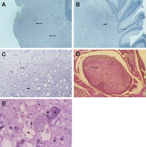
The murine leukemia virus (MLV) TR1.3 provides an excellent model to study the wide range of retrovirus-induced central nervous system (CNS) pathology and disease. TR1.3 rapidly induces thrombotic events in brain microvessels and causes cell-specific syncytium formation of brain capillary endothelial cells (BCEC). A single amino acid substitution, W102G, in the MLV envelope protein (Env) regulates the pathogenic effects. The role of Env in determining this disease phenotype compared to the induction of spongiform encephalomyelitis with a longer latency, as seen in several other MLV and in human retroviruses, was determined by studying in vitro-attenuated TR1.3. Virus cloned from this selection, termed TRM, induced progressive neurological disease characterized by ataxia and paralysis and the appearance of spongiform neurodegeneration throughout the brain stem and spinal cord. This disease was associated with virus replication in both BCEC and highly ramified glial cells. TRM did not induce syncytium formation, either in vivo or in vitro. Sequence and mutational analyses demonstrated that TRM contained a reversion of Env G102W but that neurological disease mapped to the single amino acid substitution Env S159P. The results demonstrate that single nucleotide changes within disparate regions of Env control dramatically different CNS disease patterns.
Figures


Similar articles
-
TR1.3 viral pathogenesis and syncytium formation are linked to Env-Gag cooperation.J Virol. 2007 Oct;81(19):10777-85. doi: 10.1128/JVI.00816-07. Epub 2007 Jul 18. J Virol. 2007. PMID: 17634219 Free PMC article.
-
Induction of syncytia by neuropathogenic murine leukemia viruses depends on receptor density, host cell determinants, and the intrinsic fusion potential of envelope protein.J Virol. 1999 Nov;73(11):9377-85. doi: 10.1128/JVI.73.11.9377-9385.1999. J Virol. 1999. PMID: 10516046 Free PMC article.
-
Neuropathogenic murine leukemia virus TR1.3 induces selective syncytia formation of brain capillary endothelium.Virology. 2004 Mar 30;321(1):57-64. doi: 10.1016/j.virol.2003.12.002. Virology. 2004. PMID: 15033565
-
Differential glycosylation of the Cas-Br-E env protein is associated with retrovirus-induced spongiform neurodegeneration.J Virol. 2000 Feb;74(3):1558-65. doi: 10.1128/jvi.74.3.1558-1565.2000. J Virol. 2000. PMID: 10627570 Free PMC article. Review.
-
Neurotropic retroviruses of wild mice and macaques.Ann Neurol. 1988;23 Suppl:S201-6. doi: 10.1002/ana.410230745. Ann Neurol. 1988. PMID: 3279903 Review.
Cited by
-
Misfolding of CasBrE SU is reversed by interactions with 4070A Env: implications for gammaretroviral neuropathogenesis.Retrovirology. 2010 Nov 5;7:93. doi: 10.1186/1742-4690-7-93. Retrovirology. 2010. PMID: 21054857 Free PMC article.
-
Postinhibitory rebound neurons and networks are disrupted in retrovirus-induced spongiform neurodegeneration.J Neurophysiol. 2014 Aug 1;112(3):683-704. doi: 10.1152/jn.00227.2014. Epub 2014 May 14. J Neurophysiol. 2014. PMID: 25252336 Free PMC article.
-
TR1.3 viral pathogenesis and syncytium formation are linked to Env-Gag cooperation.J Virol. 2007 Oct;81(19):10777-85. doi: 10.1128/JVI.00816-07. Epub 2007 Jul 18. J Virol. 2007. PMID: 17634219 Free PMC article.
-
Unique N-linked glycosylation of CasBrE Env influences its stability, processing, and viral infectivity but not its neurotoxicity.J Virol. 2013 Aug;87(15):8372-87. doi: 10.1128/JVI.00392-13. Epub 2013 May 22. J Virol. 2013. PMID: 23698308 Free PMC article.
-
Naturally Occurring Polymorphisms of the Mouse Gammaretrovirus Receptors CAT-1 and XPR1 Alter Virus Tropism and Pathogenicity.Adv Virol. 2011;2011:975801. doi: 10.1155/2011/975801. Epub 2011 Oct 23. Adv Virol. 2011. PMID: 22312361 Free PMC article.
References
-
- Barmak, K., E. Harhaj, C. Grant, T. Alefantis, and B. Wigdahl. 2003. Human T cell leukemia virus type I-induced disease: pathways to cancer and neurodegeneration. Virology 308:1-12. - PubMed
-
- Czub, M., S. Czub, M. Rappold, S. Mazgareanu, S. Schwender, M. Demuth, A. Hein, and R. Dorries. 1995. Murine leukemia virus-induced neurodegeneration of rats: enhancement of neuropathogenicity correlates with enhanced viral tropism for macrophages, microglia, and brain vascular cells. Virology 214:239-244. - PubMed
MeSH terms
Substances
LinkOut - more resources
Full Text Sources
