

The microphthalmia transcription factor (Mitf) controls expression of the ocular albinism type 1 gene: link between melanin synthesis and melanosome biogenesis
- PMID: 15254223
- PMCID: PMC444869
- DOI: 10.1128/MCB.24.15.6550-6559.2004
The microphthalmia transcription factor (Mitf) controls expression of the ocular albinism type 1 gene: link between melanin synthesis and melanosome biogenesis
Abstract
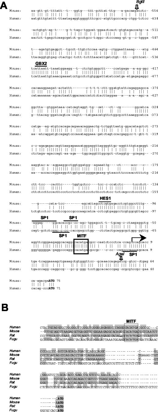
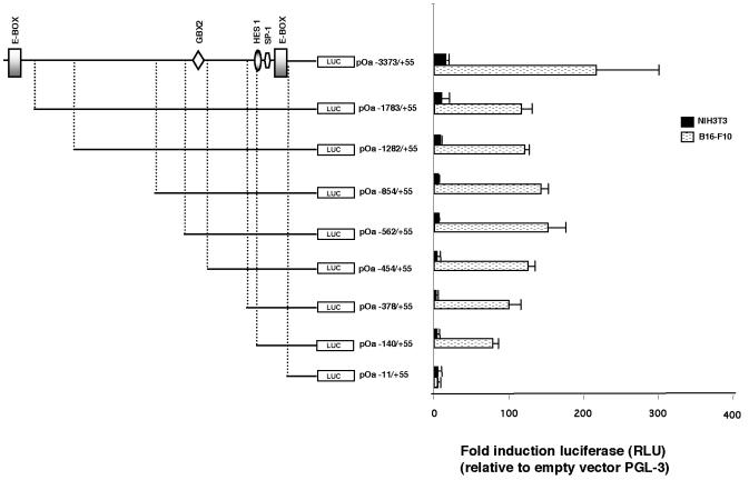
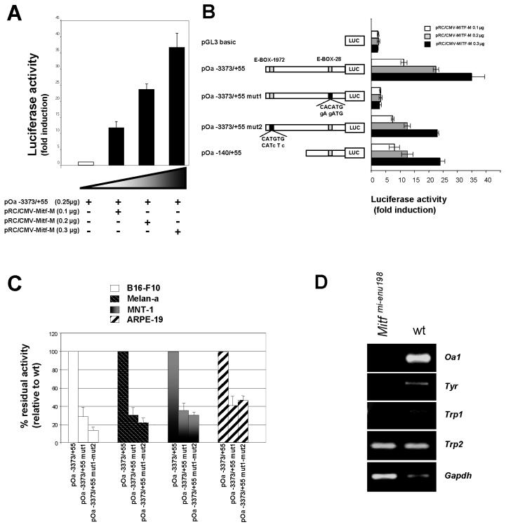
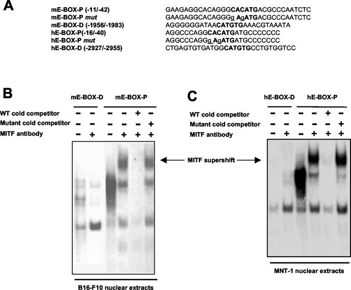
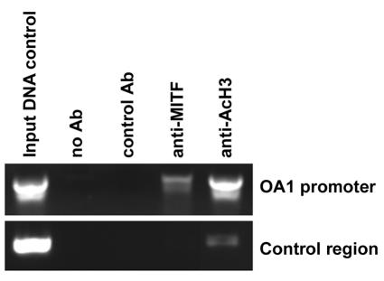
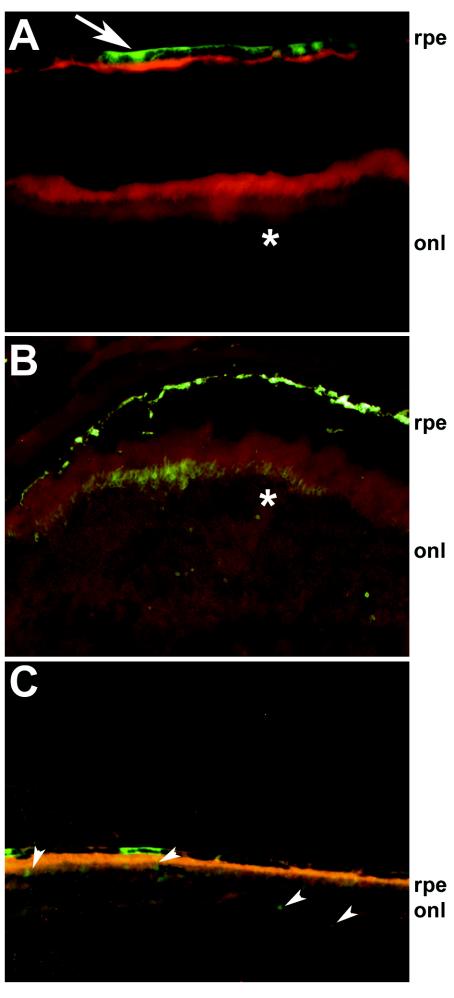
Melanogenesis is the process that regulates skin and eye pigmentation. Albinism, a genetic disease causing pigmentation defects and visual disorders, is caused by mutations in genes controlling either melanin synthesis or melanosome biogenesis. Here we show that a common transcriptional control regulates both of these processes. We performed an analysis of the regulatory region of Oa1, the murine homolog of the gene that is mutated in the X-linked form of ocular albinism, as Oa1's function affects melanosome biogenesis. We demonstrated that Oa1 is a target of Mitf and that this regulatory mechanism is conserved in the human gene. Tissue-specific control of Oa1 transcription lies within a region of 617 bp that contains the E-box bound by Mitf. Finally, we took advantage of a virus-based system to assess tissue specificity in vivo. To this end, a small fragment of the Oa1 promoter was cloned in front of a reporter gene in an adeno-associated virus. After we injected this virus into the subretinal space, we observed reporter gene expression specifically in the retinal pigment epithelium, confirming the cell-specific expression of the Oa1 promoter in the eye. The results obtained with this viral system are a preamble to the development of new gene delivery approaches for the treatment of retinal pigment epithelium defects.
Figures
Similar articles
-
Expression pattern of the ocular albinism type 1 (Oa1) gene in the murine retinal pigment epithelium.Invest Ophthalmol Vis Sci. 2000 Dec;41(13):4333-7. Invest Ophthalmol Vis Sci. 2000. PMID: 11095635
-
The ocular albinism type 1 (OA1) gene controls melanosome maturation and size.Invest Ophthalmol Vis Sci. 2005 Dec;46(12):4358-64. doi: 10.1167/iovs.05-0834. Invest Ophthalmol Vis Sci. 2005. PMID: 16303920
-
OTX2 activates the molecular network underlying retina pigment epithelium differentiation.J Biol Chem. 2003 Jun 13;278(24):21721-31. doi: 10.1074/jbc.M301708200. Epub 2003 Mar 27. J Biol Chem. 2003. PMID: 12663655
-
The ocular albinism type 1 (OA1) protein and the evidence for an intracellular signal transduction system involved in melanosome biogenesis.Pigment Cell Res. 2005 Aug;18(4):227-33. doi: 10.1111/j.1600-0749.2005.00240.x. Pigment Cell Res. 2005. PMID: 16029416 Review.
-
Ocular albinism type 1: more than meets the eye.Pigment Cell Res. 2001 Aug;14(4):243-8. doi: 10.1034/j.1600-0749.2001.140403.x. Pigment Cell Res. 2001. PMID: 11549106 Review.
Cited by
-
A molecular systems approach to modelling human skin pigmentation: identifying underlying pathways and critical components.BMC Res Notes. 2015 Apr 29;8:170. doi: 10.1186/s13104-015-1128-6. BMC Res Notes. 2015. PMID: 25925987 Free PMC article.
-
Melanocytes in regenerative medicine applications and disease modeling.J Transl Med. 2024 Apr 8;22(1):336. doi: 10.1186/s12967-024-05113-x. J Transl Med. 2024. PMID: 38589876 Free PMC article. Review.
-
Comparative transcriptome and histological analyses provide insights into the skin pigmentation in Minxian black fur sheep (Ovis aries).PeerJ. 2021 Apr 27;9:e11122. doi: 10.7717/peerj.11122. eCollection 2021. PeerJ. 2021. PMID: 33986980 Free PMC article.
-
A dual role for SOX10 in the maintenance of the postnatal melanocyte lineage and the differentiation of melanocyte stem cell progenitors.PLoS Genet. 2013;9(7):e1003644. doi: 10.1371/journal.pgen.1003644. Epub 2013 Jul 25. PLoS Genet. 2013. PMID: 23935512 Free PMC article.
-
BEST1 expression in the retinal pigment epithelium is modulated by OTX family members.Hum Mol Genet. 2009 Jan 1;18(1):128-41. doi: 10.1093/hmg/ddn323. Epub 2008 Oct 10. Hum Mol Genet. 2009. PMID: 18849347 Free PMC article.
References
-
- Amiel, J., P. M. Watkin, M. Tassabehji, A. P. Read, and R. M. Winter. 1998. Mutation of the MITF gene in albinism-deafness syndrome (Tietz syndrome). Clin. Dysmorphol. 7:17-20. - PubMed
-
- Auricchio, A., M. Hildinger, E. O'Connor, G. P. Gao, and J. M. Wilson. 2001. Isolation of highly infectious and pure adeno-associated virus type 2 vectors with a single-step gravity-flow column. Hum. Gene Ther. 12:71-76. - PubMed
-
- Auricchio, A., G. Kobinger, V. Anand, M. Hildinger, E. O'Connor, A. M. Maguire, J. M. Wilson, and J. Bennett. 2001. Exchange of surface proteins impacts on viral vector cellular specificity and transduction characteristics: the retina as a model. Hum. Mol. Genet. 10:3075-3081. - PubMed
-
- Barsh, G. S. 1996. The genetics of pigmentation: from fancy genes to complex traits. Trends Genet. 12:299-305. - PubMed
Publication types
MeSH terms
Substances
Grants and funding
LinkOut - more resources
Full Text Sources
Other Literature Sources
Molecular Biology Databases