. 2004 Aug;24(15):6620-30.
doi: 10.1128/MCB.24.15.6620-6630.2004.

Functional complementation of human centromere protein A (CENP-A) by Cse4p from Saccharomyces cerevisiae

Affiliations

Functional complementation of human centromere protein A (CENP-A) by Cse4p from Saccharomyces cerevisiae

Gerhard Wieland et al. Mol Cell Biol. 2004 Aug.

Abstract

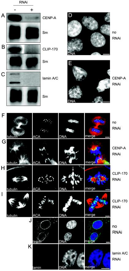
We have employed a novel in vivo approach to study the structure and function of the eukaryotic kinetochore multiprotein complex. RNA interference (RNAi) was used to block the synthesis of centromere protein A (CENP-A) and Clip-170 in human cells. By coexpression, homologous kinetochore proteins from Saccharomyces cerevisiae were then tested for the ability to complement the RNAi-induced phenotypes. Cse4p, the budding yeast CENP-A homolog, was specifically incorporated into kinetochore nucleosomes and was able to complement RNAi-induced cell cycle arrest in CENP-A-depleted human cells. Thus, Cse4p can structurally and functionally substitute for CENP-A, strongly suggesting that the basic features of centromeric chromatin are conserved between yeast and mammals. Bik1p, the budding yeast homolog of human CLIP-170, also specifically localized to kinetochores during mitosis, but Bik1p did not rescue CLIP-170 depletion-induced cell cycle arrest. Generally, the newly developed in vivo complementation assay provides a powerful new tool for studying the function and evolutionary conservation of multiprotein complexes from yeast to humans.


Figures

FIG. 1.
FIG. 1.
Budding yeast kinetochore proteins Cse4p and Bik1p are recruited to centromeres in human cells. HEp-2 cells stably expressing GFP-CENP-A (A and B), GFP-Cse4p (C and D), GFP-CLIP-170 (E to G), or GFP-Bik1p (H to J) were grown on coverslips and processed for indirect immunofluorescence for analyses of subcellular localization. Fixed cells were costained with a human anticentromere autoimmune serum to detect centromeres (ACA) and with ToPro-3 to visualize DNA. Single confocal sections were acquired from cells in interphase (i), telophase (t), or prometaphase (p). Fluorescence signals from each channel are displayed. To show the degree of colocalization, we also show overlay images of the GFP (green) and centromere signals (red). Bars, 5 μm.
FIG. 2.
FIG. 2.
GFP fusion constructs are expressed as full-length proteins. (A) Total protein lysates from HEp-2 cells (lane 1) and HEp-2 cells expressing GFP (lane 2), GFP-CENP-A (lane 3), GFP-Cse4p (lane 4), GFP-CLIP-170 (lane 5), or GFP-Bik1p (lane 6) were separated by SDS-PAGE and transferred to nitrocellulose membranes. Single strips from the membranes were probed with anticentromere autoimmune serum (lane 1) or an anti-GFP antibody (lanes 2 to 6). The ACA serum detects CENP-A at ca. 20 kDa, CENP-B at ca. 80 kDa, and CENP-C at ca. 140 kDa. GFP fusions were detected as full-length proteins at their expected molecular masses. Numbers to the right indicate the positions of standard protein markers (in kilodaltons). (B) Chromatin fractionation. DNAs extracted from equivalent supernatant (S) and pellet (P) fractions after MNase digestion were electrophoresed in a 1.5% agarose gel, followed by ethidium bromide staining (chromatin). The migration positions of mononucleosomes (mn) and polynucleosomes (pn) are indicated. Whole-cell protein extracts (WCE) and aliquots of the chromatin fractionation procedure from different HEp-2 cell lines were subjected to SDS-17.5% PAGE and Western blotting with antibodies against N-terminally acetylated histone H3 (acH3), histone H3 methylated at lysine 9 (meH3), CENP-A, and GFP, as indicated. Nucleosomes containing acetylated histone H3 or histone H3 methylated at lysine 9 were released from pellet fractions into soluble supernatant fractions with increasing amounts of MNase. In contrast, CENP-A-, GFP-CENP-A-, and GFP-Cse4p-containing nucleosomes were not released into soluble fractions under these conditions. C, cytoplasmic fraction; N, nuclear fraction.
FIG.3.
FIG.3.
CENP-A and CLIP-170 reduction by RNAi causes severe cellular defects. RNAi was used to block the protein synthesis of CENP-A (A), CLIP-170 (B), and as a control, lamin A/C (C) in HEp-2 cells. The reduction in protein level was then analyzed by immunoblotting of protein lysates from cells treated without (−) or with (+) RNAi oligonucleotides for 72 h. An anti-splicing factor SmB/B′ antibody was used as a loading control (Sm). Control cells treated without RNAi (D) and cells treated for 72 h with anti-CENP-A RNAi oligonucleotides were fixed on coverslips and stained with a DNA dye to visualize the nuclei. While the control cells had normal, round nuclei and a typical chromatin distribution throughout the nucleus during interphase (D), CENP-A-depleted cells showed smaller nuclei displaying aberrant morphologies and partial chromatin condensation (E). (F to I) The mitotic phenotypes of cells treated with RNAi against CENP-A and CLIP-170 were assessed by triple fluorescence staining of tubulin (red), centromeres (ACA [green]), and DNA (ToPro-3 [blue]). Images show single confocal sections for each staining pattern as well as overlay views (merge). While control cells showed normal spindle morphology, centromere distribution, and DNA staining during telophase (F), CENP-A-depleted cells had lagging chromosomes (G, arrows). CLIP-170-depleted mitotic cells showed misaligned and/or lagging chromosomes (H, arrows) within anaphase spindles or multipolar spindles with misaligned chromosomes (I). (J) Lamin A/C antibody staining (green) in control cells showed a typical nuclear rim pattern, while in RNAi-treated cells, lamin A/C staining was strongly diminished (K). Note that the nuclear morphology was unaffected in lamin A/C-depleted cells. Bars, 5 μm.
FIG. 4.
FIG. 4.
Inhibition of proliferation by RNAi against CENP-A and CLIP-170. (A) The frequency of mitotic cells was assessed by immunofluorescence staining of centromeres and DNA in RNAi-untreated cells (mock) or cells incubated for 72 h with RNAi oligonucleotides specific for CENP-A, CLIP-170, or lamin A/C. The diagram shows mean values with standard deviations from four independent experiments (n > 400 for each RNAi experiment). (B) Quantitation of living cells after RNAi. Identical numbers of HEp-2 cells were incubated without (mock) or with the indicated RNAi oligonucleotides for 72 h. The frequency of living cells was then determined by trypan blue exclusion. Data represent mean values and standard deviations from at least five independent experiments (n > 200 for each RNAi experiment). The values were normalized to that for mock-treated cells (set to 100%) to directly compare the effects of RNAi treatments.
FIG. 5.
FIG. 5.
Budding yeast Cse4p functionally substitutes for CENP-A in human cells. (A) Schematic representation of the assay developed to test the ability of S. cerevisiae kinetochore proteins to functionally complement centromere protein homologs in human cells. Twelve hours prior to RNAi treatment against the endogenous centromere protein, HEp-2 cells were transiently transfected with GFP expression plasmids encoding the homologous protein from budding yeast. The phenotypes of these cells were analyzed after 72 h of RNAi infection. (B) The procedure described in panel A was applied to cells that were not transfected with plasmids or RNAi treated (mock) and to CENP-A- and CLIP-170-depleted cells. The frequencies of living cells were determined at 72 h post-RNAi by trypan blue exclusion. The use of cotransfection plasmids encoding GFP fusion proteins of Cse4p, CENP-A, Bik1p, or CLIP-170 (as described in panel A) is indicated at the bottom. (C) CENP-A RNAi-treated cells cotransfected with GFP-Cse4p were fixed and immunostained to visualize DNA, centromeres, and GFP-Cse4p. The image shows individual channels of a single confocal section from a telophase cell. The Cse4p signals coincide with the positions of centromeres. (D) CLIP-170 RNAi-treated cells cotransfected with GFP-Bik1p were fixed and immunostained to visualize DNA, tubulin, and GFP-Bik1p. These cells show mitotic defects with misaligned chromosomes (upper cell) or multipolar spindles (lower cell). Bik1p is distributed throughout the whole cell, with preferred staining on chromatin. Bars, 5 μm. (E) Untreated HEp-2 cells (circles) and HEp-2 cells (open triangles) or stably Cse4-expressing HEp-2 cells (closed triangles) treated with anti-CENP-A RNAi were observed for 3 days post-RNAi oligonucleotide administration. The frequencies of living cells were determined for at least three independent experiments at 0, 24, 48, and 72 h. While the proliferation of HEp-2 cells was significantly reduced by CENP-A RNAi, the HEp-2/Cse4 cell line proliferated with kinetics similar to those of mock-treated HEp-2 cells.

Similar articles

Cited by

References

    1. Albertson, D. G., and J. N. Thomson. 1982. The kinetochores of Caenorhabditis elegans. Chromosoma 86:409-428. - PubMed
    1. Albertson, D. G., and J. N. Thomson. 1993. Segregation of holocentric chromosomes at meiosis in the nematode, Caenorhabditis elegans. Chromosome Res. 1:15-26. - PubMed
    1. Amon, A. 1999. The spindle checkpoint. Curr. Opin. Genet. Dev. 9:69-75. - PubMed
    1. Arents, G., R. W. Burlingame, B. C. Wang, W. E. Love, and E. N. Moudrianakis. 1991. The nucleosomal core histone octamer at 3.1 A resolution: a tripartite protein assembly and a left-handed superhelix. Proc. Natl. Acad. Sci. USA 88:10148-10152. - PMC - PubMed
    1. Biggins, S., and A. W. Murray. 1999. Sister chromatid cohesion in mitosis. Curr. Opin. Genet. Dev. 9:230-236. - PubMed

MeSH terms

LinkOut - more resources