. 2004 Aug;24(15):6719-27.
doi: 10.1128/MCB.24.15.6719-6727.2004.

Gene-targeted mice lacking the Trex1 (DNase III) 3'-->5' DNA exonuclease develop inflammatory myocarditis

Affiliations

Gene-targeted mice lacking the Trex1 (DNase III) 3'-->5' DNA exonuclease develop inflammatory myocarditis

Masashi Morita et al. Mol Cell Biol. 2004 Aug.

Abstract

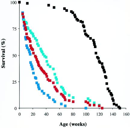
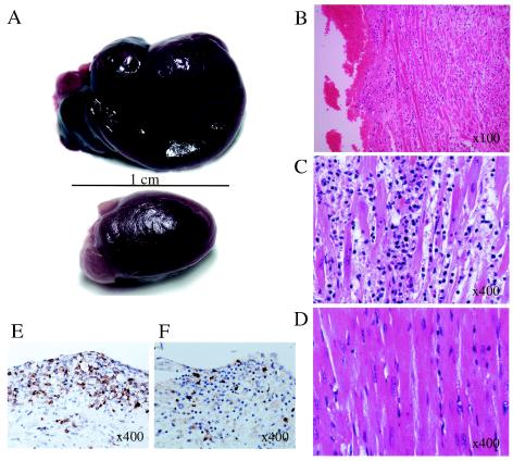
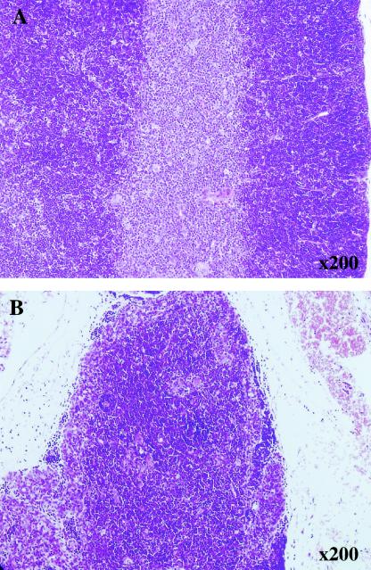
TREX1, originally designated DNase III, was isolated as a major nuclear DNA-specific 3'-->5' exonuclease that is widely distributed in both proliferating and nonproliferating mammalian tissues. The cognate cDNA shows homology to the editing subunit of the Escherichia coli replicative DNA polymerase III holoenzyme and encodes an exonuclease which was able to serve a DNA-editing function in vitro, promoting rejoining of a 3' mismatched residue in a reconstituted DNA base excision repair system. Here we report the generation of gene-targeted Trex1(-/-) mice. The null mice are viable and do not show the increase in spontaneous mutation frequency or cancer incidence that would be predicted if Trex1 served an obligatory role of editing mismatched 3' termini generated during DNA repair or DNA replication in vivo. Unexpectedly, Trex1(-/-) mice exhibit a dramatically reduced survival and develop inflammatory myocarditis leading to progressive, often dilated, cardiomyopathy and circulatory failure.


Figures

FIG. 1.
FIG. 1.
Targeted disruption of murine Trex1 locus. (A) Physical map of genomic DNA containing the Trex1 gene. Genomic sequences are shown as a solid line, with the single Trex1 coding exon depicted as an open box and the three conserved exonuclease motifs indicated by vertical black bars within it; vector sequences are shown with dashed lines. Genomic DNA fragments were subcloned on either side of a neo cassette to generate a construct that deleted sequences encoding amino acids 103 to 207 of the 304-amino-acid Trex1 ORF product (including motifs II and III) at the targeted locus. Restriction digestion with BamHI gave rise to a 3.6-kb fragment at the wild-type locus and a 2.8-kb fragment at the targeted locus that were detected by hybridization with a 3′ flanking probe (filled box). The positions of sense and antisense PCR primers 1 and 2, which amplify a 0.5-kb wild-type product and a 1.3-kb product from the targeted locus, are indicated. Genotyping of representative wild-type (+/+), heterozygous (+/−), and Trex1 null (−/−) live-born F2 progeny from tail-snip biopsy samples by Southern hybridization analysis with the 3′ probe (B) or by PCRs with primers 1 and 2 (C) is shown. (D) RT-PCR analysis of increasing amounts of total RNA (5, 50, and 500 ng) from spleens of wild-type (+/+) and Trex1 null (−/−) mice. A Trex1-specific signal (arrow; same primers as for panel A) was detected for the wild-type but not the Trex1 null mice (left); a signal for the glyceraldehyde-3-phosphate dehydrogenase control (see Materials and Methods) was readily detected in both wild-type and null samples (right). The migration of DNA markers (in kilobases) is shown. (E) Immunoblot analysis of MEF (left) and liver (right) nuclear protein extracts from wild-type (+/+) and Trex1 null (−/−) mice with a murine Trex1-specific antipeptide antiserum. The migration of full-length Trex1 is indicated in each case (arrows); a Trex1 fragment for the wild-type liver is marked by an asterisk. The migration of molecular mass markers (in kilodaltons) is indicated.
FIG. 2.
FIG. 2.
DNA 3′ exonuclease activity in nuclear extracts of Trex1−/− and Trex1+/+ cells and tissues. Trex1−/− (open boxes) and Trex1+/+ (filled boxes) crude nuclear protein extracts from livers (A), testes (B), and thymus glands (C) were assayed for 3′ exonuclease activity with a 3′-end-labeled poly(dA) substrate. (D) Extracts from Trex1−/− (−/−) and Trex1+/+ (+/+) MEF cell lines were assayed before and after partial purification by column chromatography on single-stranded DNA (ssDNA)-cellulose, as indicated. (E) An extract of Trex1+/+ mouse liver was loaded onto a single-stranded DNA-cellulose column and eluted with increasing salt concentrations (as indicated). The protein fractions were quantified (open circles) and assayed as described above for 3′ exonuclease activity (filled boxes) and by immunoblotting with a specific antiserum which detects full-length Trex1 (arrow) and a proteolytic fragment (asterisk) (as in Fig. 1E).
FIG. 3.
FIG. 3.
Reduced survival of Trex1 null mice. Survival curves are shown for all Trex1−/− mice in this study (n = 110; red curve) and for the wild-type Trex1+/+ controls (n = 77; black curve). For those Trex1−/− mice that were born to heterozygous (n = 51; dark blue curve) or homozygous null (n = 59; light blue curve) mothers, the difference in median survival of 21 weeks was significant (log rank test with 95% confidence interval; P < 0.0001).
FIG. 4.
FIG. 4.
Heart pathology in Trex1 null mice. (A) Auricular thrombus and distended heart in a Trex1−/− female mouse that died of circulatory failure at 40 weeks; the heart from a female control sacrificed at the same age is shown for comparison below. (B and C) Section through left auricle of affected 10-week-old animal (hematoxylin-eosin staining). (B) The normal myocardial muscle pattern is disrupted, with the red fibers being separated by a cellular inflammatory infiltrate with edema. The endocardial surface has a layer of adherent thrombus (left side of field). (C) At a higher magnification, the cardiac fibers appear to be degenerate, with a mixed infiltrate of dark lymphoid cells and polymorphs; (D) a normal control is shown for comparison. (E and F) Immunostaining of infiltrating lymphoid cells in a Trex1−/− heart shows predominantly CD3-positive T cells in the myocardium and the subendocardial region (E), with some CD45R-expressing B cells present as mainly perivascular aggregates in the subendocardial region (F); relatively few morphologically identifiable plasma cells were present. A scale bar and original magnifications are shown.
FIG. 5.
FIG. 5.
Thymus pathology in Trex1 null mice. (A) Normal thymus from a Trex1+/+ control shows clear distinction between the darker lymphocyte-rich cortex and the paler medulla. (B) Atrophic thymus from a representative Trex1−/− mouse has indistinct corticomedullary junction and is smaller overall. The mice were females 10 to 12 weeks old. Original magnifications are indicated.

Similar articles

Cited by

References

    1. Bachmaier, K., J. Le, and J. M. Penninger. 2000. “Catching heart disease”: antigenic mimicry and bacterial infections. Nat. Med. 6:841-842. - PubMed
    1. Bennett, R. A., D. M. Wilson, D. Wong, and B. Demple. 1997. Interaction of apurinic endonuclease and DNA polymerase β in the base excision repair pathway. Proc. Natl. Acad. Sci. USA 94:7166-7169. - PMC - PubMed
    1. Brown, K. R., K. L. Weatherdon, C. L. Galligan, and V. Skalski. 2002. A nuclear 3′-5′ exonuclease proofreads for the exonuclease-deficient DNA polymerase α. DNA Repair 1:795-810. - PubMed
    1. Chen, J., R. Lansford, V. Stewart, F. Young, and F. W. Alt. 1993. RAG-2-deficient blastocyst complementation: an assay of gene function in lymphocyte development. Proc. Natl. Acad. Sci. USA 90:4528-4532. - PMC - PubMed
    1. Chou, K.-M., and Y.-C. Cheng. 2002. An exonucleolytic activity of human apurinic/apyrimidinic endonuclease on 3′ mispaired DNA. Nature 415:655-659. - PubMed

Publication types

MeSH terms