. 2004 Jul;114(2):172-81.
doi: 10.1172/JCI20641.

Evidence for a critical contribution of haploinsufficiency in the complex pathogenesis of Marfan syndrome

Affiliations

Evidence for a critical contribution of haploinsufficiency in the complex pathogenesis of Marfan syndrome

Daniel P Judge et al. J Clin Invest. 2004 Jul.

Abstract

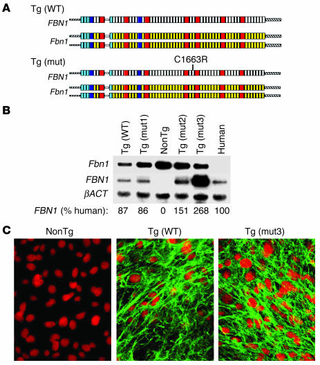
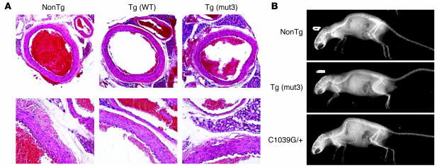
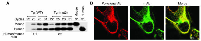
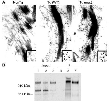
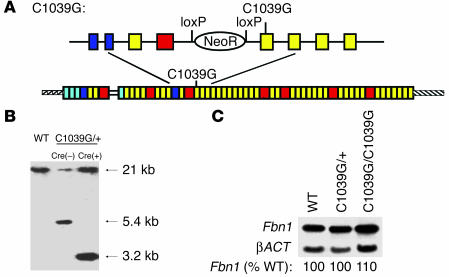
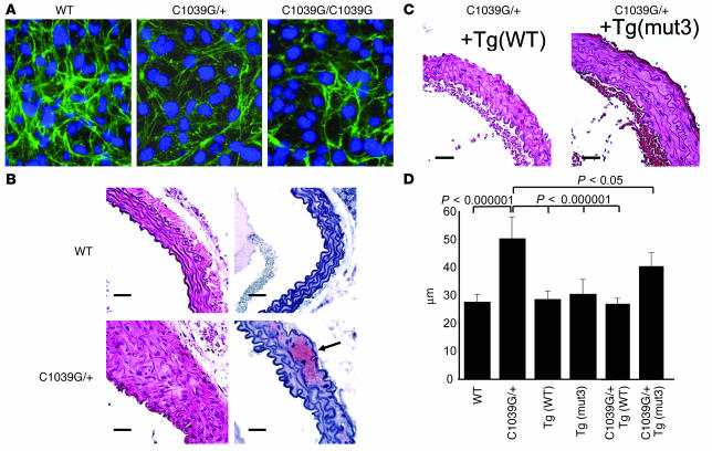
Marfan syndrome is a connective tissue disorder caused by mutations in the gene encoding fibrillin-1 (FBN1). A dominant-negative mechanism has been inferred based upon dominant inheritance, mulitimerization of monomers to form microfibrils, and the dramatic paucity of matrix-incorporated fibrillin-1 seen in heterozygous patient samples. Yeast artificial chromosome-based transgenesis was used to overexpress a disease-associated mutant form of human fibrillin-1 (C1663R) on a normal mouse background. Remarkably, these mice failed to show any abnormalities of cellular or clinical phenotype despite regulated overexpression of mutant protein in relevant tissues and developmental stages and direct evidence that mouse and human fibrillin-1 interact with high efficiency. Immunostaining with a human-specific mAb provides what we believe to be the first demonstration that mutant fibrillin-1 can participate in productive microfibrillar assembly. Informatively, use of homologous recombination to generate mice heterozygous for a comparable missense mutation (C1039G) revealed impaired microfibrillar deposition, skeletal deformity, and progressive deterioration of aortic wall architecture, comparable to characteristics of the human condition. These data are consistent with a model that invokes haploinsufficiency for WT fibrillin-1, rather than production of mutant protein, as the primary determinant of failed microfibrillar assembly. In keeping with this model, introduction of a WT FBN1 transgene on a heterozygous C1039G background rescues aortic phenotype.


Figures

Figure 1
Figure 1
(A) Schematic representation of YAC transgenics. The endogenous untargeted mouse alleles (Fbn1) are shown with full-length WT human FBN1 or human FBN1 harboring a mutation (mut) that results in classic MFS in humans (C1663R). The cbEGF-like domains are shown in yellow (mouse) or white (human); 8-cys LTBP-binding domains are shown in red, and hybrid domains are shown in blue. (B) Northern blot analysis was performed on cultured dermal fibroblasts isolated from each of the transgenic strains. The radiolabeled probes recognize the 3′ UTRs of Fbn1 or FBN1. Expression of the genes encoding fibrillin-1 is normalized to expression of the gene encoding β-actin (βACT) and compared with the amount of FBN1 expression in a human fibroblast cell line isolated from a WT control individual. NonTg, nontransgenic. (C) Cultured dermal fibroblasts stained with mAb to human fibrillin-1. The left panel is from a littermate nontransgenic mouse; Tg(WT) is from a mouse with YAC containing WT FBN1; Tg(mut3) is from a mouse with YAC containing mutant FBN1 (C1663R).
Figure 2
Figure 2
(A) Representative aortic wall sections from mice aged 12 months, stained with H&E. NonTg is the nontransgenic control, Tg(WT) is from the strain expressing WT FBN1, and Tg(mut3) is from the strain with the highest expression of C1663R-mutant FBN1. Magnification, ×10 (top) and ×40 (bottom). In the low-power views, the adjacent pulmonary artery is visible. No differences were detected. (B) Representative radiographs taken of mice at 1 year of age. Overgrowth of the ribs and kyphoscoliosis are evident in the C1039G heterozygote (C1039G/+) mice (bottom), without discernible bony abnormalities in the Tg(mut3) mice, in comparison with nonTg control mice.
Figure 3
Figure 3
(A) Relative quantification of YAC transgene expression compared with endogenous Fbn1 expression. RT-PCR amplicons were generated using total RNA isolated from mouse aorta and oligonucleotides complementary to exons 10 and 11 that recognize identical sequence in mice and humans. The amplicon from FBN1 has a unique TaqI digest site, resulting in a smaller band that binds to a probe generated from 32P-labeled antisense oligonucleotide in exon 11. Ratio of human to mouse mRNA in the exponential portion of amplification is indicated below. Tg(mut3) is the highest expressing strain, while two other strains were intermediate in their FBN1 expression. (B) Developing limb from Tg(mut3) mouse stained for fibrillin-1, demonstrating robust transgenic production of mutant human fibrillin-1 in tissues expressing endogenous mouse fibrillin-1. Left panel: polyclonal Ab that detects both mouse and human fibrillin-1; middle panel: mAb that is specific to human fibrillin-1; right panel: overlay.
Figure 4
Figure 4
(A) Immunoelectron microscopy of mouse skin using an mAb that is specific for human fibrillin-1. Left panel, nontransgenic (NonTg) control, shows no staining, confirming previously described specificity of this Ab. Middle panel, Tg(WT), and right panel, Tg(mut3), both show Ab recognition of human fibrillin-1 in a pattern that suggests the presence of both murine and human proteins within the same microfibrillar bundle. Box in the lower-right corner highlights the pattern of epitope staining, seen in black. (B) Murine and human fibrillin-1 interact. Lanes 1 and 4: 35S-labeled media from murine fibroblasts coincubated with unlabeled media from murine fibroblasts before and after immunoprecipitation (IP), respectively; lanes 2 and 5: identical treatment of 35S-labeled media from murine fibroblasts coincubated with unlabeled media from human fibroblasts before and after IP, respectively; lanes 3 and 6: identical treatment of 35S-labeled media from human fibroblasts coincubated with unlabeled media from human fibroblasts before and after IP, respectively. Results in lane 5 demonstrate interaction between 35S-labeled murine fibrillin-1 and unlabeled human fibrillin-1, while results in lane 4 attest to the specificity of the mAb for human fibrillin-1. Lane 6 serves as a positive control for IP. Position of molecular weight standards is indicated on the left.
Figure 5
Figure 5
(A) Drawing depicting cDNA resulting from proper homologous recombination of the endogenous Fbn1 allele with the targeting vector for the C1039G mutation. Above is a representation of the genomic segment prior to Cre-mediated recombination to remove the NeoR flanked by loxP sequences. (B) Representative Southern blot demonstrating correct targeting before and after Cre-mediated loxP recombination. Left lane is from a WT, untargeted mouse, with an undigested 21-kb KpnI fragment; middle lane is from a C1039G heterozygous mutant mouse without Cre-recombinase, resulting in an untargeted band (21 kb) and a targeted band (5.4 kb); right lane is from a C1039G heterozygous mutant mouse after Cre-mediated recombination, resulting in an untargeted band (21 kb) and a targeted band (3.2 kb). (C) Northern blot demonstrating that the level of expression of Fbn1 in mice heterozygous or homozygous for the C1039G allele is unchanged in comparison with untargeted (WT) mice. Expression is normalized to expression of the gene encoding β-actin (βACT). The level of Fbn1 expression in heterozygous and homozygous targeted mice, relative to that seen in WT, is indicated.
Figure 6
Figure 6
(A) Cysteine substitution in fibrillin-1 results in impaired microfibril formation. Cultured dermal fibroblasts from WT, heterozygous (C1039G/+), or homozygous (C1039G/C1039G) mice (for the Fbn1 mutation) were plated at 5 × 10⁵/ml and immunostained for fibrillin-1 using polyclonal anti–fibrillin-1 Ab 9543 after 72 hours of confluence. The quantity and quality of the microfibrils containing the product of the mutant allele appears diminished. (B) Representative aortic wall sections from mice aged 6 months, stained with H&E (left) and Safranin-O (right). Top panels are from a WT control, and bottom panels are from a mouse heterozygous for the C1039G mutation (C1039G/+). Mutant mice have focal areas of medial thickening with disorganization and disruption of elastic lamellae, as well as frequent areas of proteoglycan deposition (arrow). Magnification, ×40 for all sections. Scale bar: 25 μm. (C) Representative aortic wall sections from mice heterozygous for the C1039G mutation and harboring the WT FBN1 transgene, +Tg(WT), or the C1663R-mutant FBN1 transgene, +Tg(mut3). Aortic wall organization is greatly improved by the WT transgene and partly improved by the mutant transgene. Magnification, ×40 for all sections. Scale bar: 25 μm. (D) Average aortic wall thickness (μm) from at least five mice of each genotype at age 6 months. For each mouse, at least six independent sections were examined, and for each section, four measures of the aortic wall thickness were averaged, including the thickest and thinnest, for each section.

Comment in

References

    1. Pyeritz, R.E. 1997. Marfan syndrome and other disorders of fibrillin. In Principles and practice of medical genetics. D.L. Rimoin, J.M. Connor, and R.E. Pyeritz, editors. Churchill Livingstone. New York, New York, USA
    1. Gray JR, et al. Ascertainment and severity of Marfan syndrome in a Scottish population. J. Med. Genet. 1994;31:51–54. - PMC - PubMed
    1. Silverman DI, et al. Life expectancy in the Marfan syndrome. Am. J. Cardiol. 1995;75:157–160. - PubMed
    1. Dietz HC, et al. Marfan syndrome caused by a recurrent de novo missense mutation in the fibrillin gene. Nature. 1991;352:337–339. - PubMed
    1. Dietz HC, et al. The question of heterogeneity in Marfan syndrome. Nat. Genet. 1995;9:228–231. - PubMed

Publication types

MeSH terms