. 2004 Nov 1;383(Pt. 3):507-15.
doi: 10.1042/BJ20040175.

Purified recombinant human prosaposin forms oligomers that bind procathepsin D and affect its autoactivation

Affiliations

Purified recombinant human prosaposin forms oligomers that bind procathepsin D and affect its autoactivation

Madanan Madathiparambil Gopalakrishnan et al. Biochem J. .

Abstract

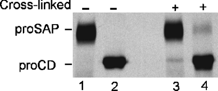
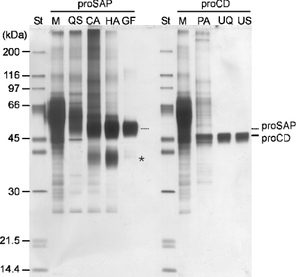
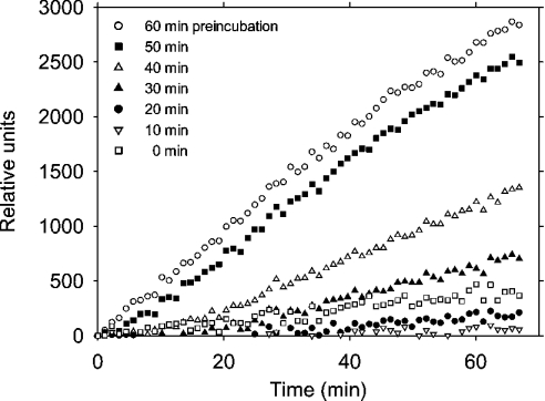
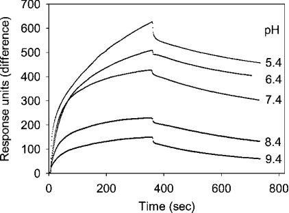
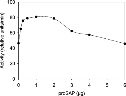
Before delivery to endosomes, portions of proCD (procathepsin D) and proSAP (prosaposin) are assembled into complexes. We demonstrate that such complexes are also present in secretions of cultured cells. To study the formation and properties of the complexes, we purified proCD and proSAP from culture media of Spodoptera frugiperda cells that were infected with baculoviruses bearing the respective cDNAs. The biological activity of proCD was demonstrated by its pH-dependent autoactivation to pseudocathepsin D and that of proSAP was demonstrated by feeding to saposin-deficient cultured cells that corrected the storage of radioactive glycolipids. In gel filtration, proSAP behaved as an oligomer and proCD as a monomer. ProSAP altered the elution of proCD such that the latter was shifted into proSAP-containing fractions. ProSAP did not change the elution of mature cathepsin D. Using surface plasmon resonance and an immobilized biotinylated proCD, binding of proSAP was demonstrated under neutral and weakly acidic conditions. At pH 6.8, specific binding appeared to involve more than one binding site on a proSAP oligomer. The dissociation of the first site was characterized by a K(D1) of 5.8+/-2.9x10(-8) M(-1) (calculated for the monomer). ProSAP stimulated the autoactivation of proCD and also the activity of pseudocathepsin D. Concomitant with the activation, proSAP behaved as a substrate yielding tri- and disaposins and smaller fragments. Our results demonstrate that proSAP forms oligomers that are capable of binding proCD spontaneously and independent of the mammalian type N-glycosylation but not capable of binding mature cathepsin D. In addition to binding proSAP, proCD behaves as an autoactivable and processing enzyme and its binding partner as an activator and substrate.


Figures

Figure 1
Figure 1. Presence of cross-linkable proSAP–proCD complexes in secretions of PMA-treated U937 cells
U937 cells were metabolically labelled in the presence of Tran35S-label, 10 mM NH4Cl and 0.2 μM PMA for 15 h. Aliquots of the medium from 6×10⁷ cells were either left untreated (–) or incubated with 1 mM dithio-bis-succinimidyl propionate cross-linking reagent for 5 min at room temperature (+) and processed for immunoprecipitation with anti-saposin C (lanes 1 and 3) and anti-proCD antibodies (lanes 2 and 4). The labelled precursors were separated by SDS/PAGE and visualized by fluorography.
Figure 2
Figure 2. Qualitative characterization of protein enrichment through purification steps of proSAP and proCD
Standard proteins and aliquots of the starting media and pools obtained through successive purification steps as indicated in the Figure were heated in the presence of SDS and dithiothreitol and subjected to SDS/PAGE. Staining of the protein with a silver nitrate method indicated that albumin was the major constituent in the starting material, whereas the purification resulted in a gradual enrichment of the recombinant proteins proSAP and proCD. ‘St’ refers to a protein standard mixture. Lane M denotes the medium and the other symbols refer to the purification steps using Q-Sepharose (QS), concanavalin A–Sepharose (CA), hydroxyapatite (HA), gel filtration (GF), pepstatin A-affinity chromatography (PA), UNO-Q ion-exchange chromatography (UQ) and UNO-S-1R ion-exchange chromatography (US) as described in the Materials and methods section. The asterisk indicates the position of trisaposin-like fragments that accompany proSAP. The standards (St), in the order of decreasing apparent masses, were myosin, β-galactosidase, phosphorylase b, BSA, ovalbumin, carbonic anhydrase, soya-bean trypsin inhibitor and lysozyme.
Figure 3
Figure 3. Stimulation of the degradation of glycosphingolipids in saposin-deficient fibroblasts with recombinant proSAP added to the medium
Control (Co) and proSAP-deficient fibroblasts (proSAP–/–) were incubated with [14C]serine (1 μCi/ml) for 24 h (pulse) followed by a chase phase of 120 h. During the pulse and chase periods, the culture medium in one proSAP(–/–) culture dish was supplemented with 25 μg/ml recombinant proSAP. Cells were harvested, and lipids were extracted and separated into acidic and neutral fractions. After chromatography, the lipids were visualized as described in the Materials and methods section. The positions of ceramide (Cer), glucosylceramide (GlcCer), lactosylceramide (LacCer), globotriaosylceramide (Globo3), globotetraosylceramide (Globo4), sphingomyelin (SM), monosialogangliosides 1–3 (GM1–GM3), disialoganglioside (GD3) and fatty acids (FA) are indicated.
Figure 4
Figure 4. Autocatalytic processing of recombinant proCD at acidic pH
ProCD (100 ng) was either left untreated (lane 0) or incubated for 10–320 min as indicated in the presence of 50 mM each of acetate and formiate/NaOH buffers (pH 3.65) in 5 μl at 37 °C. The reaction was terminated by adding 1 μl of 0.5 M Tris/HCl buffer (pH 8.0) containing 10 μM pepstatin A. The samples were denatured, deglycosylated with peptide N-glycosidase F and analysed by SDS/PAGE and silver nitrate staining.
Figure 5
Figure 5. Autocatalytic activation of recombinant proCD at acidic pH
Aliquots of proCD were incubated at pH 3.65 and 37 °C as described in the legend to Figure 4. After incubation for the periods indicated, the samples were assayed for cathepsin D activity and fluorescence was recorded under standard conditions. Untreated control and the samples that were incubated in the acidic buffer for 10, 20, 30, 40, 50 and 60 min are shown, as indicated in the plot.
Figure 6
Figure 6. Demonstration of complex formation between proSAP and proCD by gel-filtration analysis
ProSAP, proCD and their mixtures were incubated in 0.1 ml of PBS (pH 6.8) with 0.1 mM leupeptin for 1 h at room temperature and for 16 h at 4 °C. The samples were fractionated on a Superdex 200 FPLC gel-filtration column and fractions (0.25 ml each) were collected. The proteins were precipitated by mixing with 1 ml of cold acetone. The precipitates were collected by centrifugation and analysed by SDS/PAGE and silver nitrate staining of protein. In (AD), four different runs with proCD or proSAP alone and their combinations are shown. These were performed with 0/1, 0.7/1, 1.4/1 and 1.4/0 nmol of proSAP/nmol of proCD respectively. In (E), separation of a mixture of 0.7 nmol each of proSAP and placental cathepsin D (preincubated as above at pH 6.8 in the presence of 0.1 mM leupeptin and 2 μM pepstatin A) is shown. The positions of single-chain cathepsin D (scCD) and of large subunit of mature double-chain (lmCD) cathepsin D are indicated. In the left margin lane, ovalbumin (Mr 45000) and carbonic anhydrase (Mr 30000) standards are shown. The elution of Dextran Blue (Mr 2000000), β-amylase (Mr 200000), BSA (Mr 66000), carbonic anhydrase (Mr 30000) and cytochrome c (Mr 12400), which was examined separately, is indicated by arrows that refer to Mr in thousands.
Figure 7
Figure 7. Binding of proSAP to immobilized proCD at various pH values
Biotinylated proCD was adsorbed on a streptavidin–dextran-coated chip and surface plasmon resonance was recorded for 6 min with solutions of proSAP (730 nM monomer) at pH values from 5.4 to 9.4 as indicated in the Figure, and with the corresponding buffers without proSAP for another 6 min.
Figure 8
Figure 8. Binding and desorption of proSAP at different concentrations to biotinylated proCD on the surface of a streptavidin–dextran-coated chip
The reactions were examined with 1.47, 0.73, 0.37 and 0.24 μM proSAP (concentrations refer to those of the monomer) as indicated in the plot. Surface plasmon resonance in the presence and absence of proSAP was recorded as described in the legend to Figure 6, but all measurements were performed at pH 6.8. The best-fit (indicated by the dotted lines) was obtained by using the bivalent analyte model for proSAP.
Figure 9
Figure 9. Processing of proCD in the presence of proSAP after the acidification of the incubation medium
Four groups of samples containing 0.2 μg of proCD and 0.1, 0.2 and 0.4 μg of proSAP or either precursor alone (0.2 and 0.4 μg respectively) in 4 μl of PBS (pH 6.8) were incubated for 1 h at room temperature. After incubation for 0, 1, 2 or 3 h with 1 μl of 0.25 M acetate and 0.25 M sodium formate buffer (pH 3.65), the reactions were terminated by adding 1 vol. of 0.1 M Tris/HCl buffer (pH 8.0) containing 10 μM pepstatin A. The polypeptides were precipitated with 80% (v/v) acetone and solubilized, deglycosylated using peptide N-glycosidase F (PNGase F) and analysed by SDS/PAGE and silver nitrate staining. Notice the acceleration by proSAP in the processing of proCD that is apparent from the relative intensities of the proCD and pseudoCD bands. This effect was reproduced in all experiments performed (n=3).
Figure 10
Figure 10. Activation of pseudoCD in the presence of proSAP
PseudoCD was prepared by incubating proCD at pH 3.65 with a conversion of 95% of the precursor. Aliquots (100 ng each) were incubated with the indicated amounts of proSAP in 10 mM sodium phosphate buffer (pH 6.8) at room temperature for 30 min. The solutions were then mixed with the fluorescent haemoglobin substrate under standard cathepsin D assay conditions to determine the enzyme activity.
Figure 11
Figure 11. Processing of proSAP in the presence of proCD
(A) Samples containing 0.2, 0.4 or 0.8 μg of proSAP were incubated without or with up to 80 ng of proCD in 2 μl of 10 mM sodium phosphate buffer (pH 6.8) for 1 h at room temperature. After adding 2 μl of 0.25 M acetate and 0.25 M sodium formiate buffer (pH 3.65), the incubation was continued for 14 h at 37 °C. The reactions were terminated and the polypeptide fragments were analysed as described in the legend to Figure 9. (B) Mixtures of the two precursors were incubated at pH 3.65 for 18 h and analysed by Western blotting using antisera against saposins A–D as indicated in the Figure. Incubations were performed with 200 ng of proSAP and 20 ng of proCD (lanes 1), 400 ng of proSAP and 40 ng of proCD (lanes 2) and 400 ng of proSAP and 160 ng of proCD (lanes 3). The protein standards shown include aprotinin (6.5 kDa).

Similar articles

Cited by

References

    1. Qi X., Grabowski G. A. Differential membrane interactions of saposins A and C: implications for the functional specificity. J. Biol. Chem. 2001;276:27010–27017. - PubMed
    1. Sandhoff K., Kolter T. Biosynthesis and degradation of mammalian glycosphingolipids. Philos. Trans. R. Soc. Lond. B. 2003;358:847–861. - PMC - PubMed
    1. Winau F., Schwierzeck V., Hurwitz R., Remmel N., Sieling P. A., Modlin R. L., Porcelli S. A., Brinkmann V., Sugita M., Sandhoff K., et al. Saposin C is required for lipid presentation by human CD1b. Nature Immunol. 2004;5:169–174. - PubMed
    1. Hazkani-Covo E., Altman N., Horowitz M., Graur D. The evolutionary history of prosaposin: two successive tandem-duplication events gave rise to the four saposin domains in vertebrates. J. Mol. Evol. 2002;54:30–34. - PubMed
    1. Ahn V. E., Faull K. F., Whitelegge J. P., Fluharty A. L., Privé G. G. Crystal structure of saposin B reveals a dimeric shell for lipid binding. Proc. Natl. Acad. Sci. U.S.A. 2003;100:38–43. - PMC - PubMed

Publication types

MeSH terms