. 2004 Aug;53(3):857-69.
doi: 10.1111/j.1365-2958.2004.04155.x.

Cyclic diguanylate (c-di-GMP) regulates Vibrio cholerae biofilm formation

Affiliations

Cyclic diguanylate (c-di-GMP) regulates Vibrio cholerae biofilm formation

Anna D Tischler et al. Mol Microbiol. 2004 Aug.

Abstract

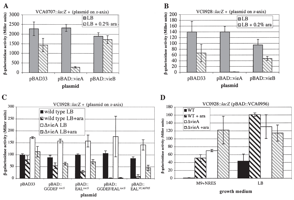
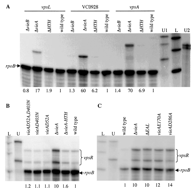
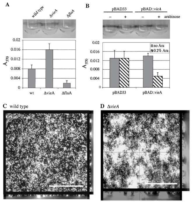
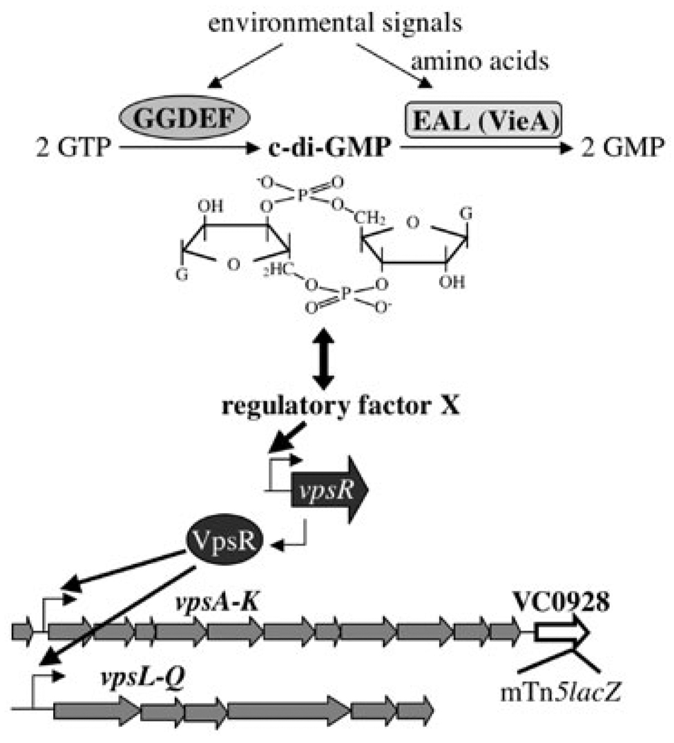
While studying virulence gene regulation in Vibrio cholerae during infection of the host small intestine, we identified VieA as a two-component response regulator that contributes to activating expression of cholera toxin. Here we report that VieA represses transcription of Vibrio exopolysaccharide synthesis (vps) genes involved in biofilm formation by a mechanism independent of its phosphorelay and DNA-binding activities. VieA controls the intracellular concentration of the cyclic nucleotide second messenger cyclic diguanylate (c-di-GMP) using an EAL domain that functions as a c-di-GMP phosphodiesterase. Two-dimensional thin layer chromatography of nucleotide extracts confirmed that VieA reduces the concentration of c-di-GMP, opposing the action of c-di-GMP synthetase proteins. Expression of unrelated V. cholerae c-di-GMP synthetase or phosphodiesterae proteins also modulated c-di-GMP concentration and vps gene expression. We propose that c-di-GMP synthetase and phosphodiesterase domain-containing proteins contribute to regulating biofilm formation by controlling c-di-GMP concentration.


Figures

Fig. 1
Fig. 1
VieA represses activity of mTn5lacZ fusions to VCA0707 and VC0928. β-Galactosidase assays were performed after overnight growth in LB (solid bars) or LB + 0.2% arabinose (hatched bars) at 30°C with shaking. Results are the average ± standard deviation for three independent cultures. A. VCA0707::lacZ activity in V. cholerae harbouring the indicated plasmids. B. VC0928::lacZ activity in V. cholerae harbouring the indicated plasmids. C. VC0928::lacZ activity in wild type and ΔvieA V. cholerae harbouring plasmids with the arabinose-inducible protein domains indicated on the x-axis. D. VC0928::lacZ activity in wild type and ΔvieA V. cholerae harbouring a plasmid encoding arabinose-inducible VCA0956. Assays were performed after overnight growth in LB ± 0.2% arabinose, or M9 + NRES ± 0.2% arabinose.
Fig. 2
Fig. 2
Ribonuclease protection assays (RPAs) to analyse regulation of vpsA vpsL, VC0928, and vpsR by VieA. RNA was isolated from wild type or the mutant V. cholerae strains indicated above each lane after growth in M9 + NRES. Transcripts were detected in 1 µg of total RNA by RPA. Band intensities were quantified and normalized to the intensity of the rpoB band. L indicates RNA ladder (sizes in number of bases = 400, 300 and 200). A. Average intensity relative to the wild type from at least three independent experiments is reported below each lane. U1 indicates undigested rpoB (bottom band), vpsA (top band), and VC0928 (middle band) probes. U2 indicates undigested vpsL probe (lower band) and an unrelated probe. B and C. The intensity of the full-length (top) vpsR protected band relative to the wild type for at least three independent experiments is reported below each lane. U indicates uncut vpsR and rpoB probes.
Fig. 3
Fig. 3
Assays for biofilm formation. A. The wild type, ΔvieA and ΔflaA strains were grown in LB without aeration and adherent bacteria were stained with crystal violet. Staining was quantified by solubilizing crystal violet in 100% ethanol and determining the absorbance at 570 nm. B. Crystal violet staining and quantitation was performed as in part A for the wild type strain harbouring either pBAD33 or pBAD33::vieA grown in LB ± 0.2% arabinose. C and D. Confocal fluorescence microscopy images of wild type and ΔvieA biofilms grown on coverslips and stained with DAPI. The centre of each figure is an XY-section through the bio-film (bar = 25 µm). Vertical sections through the biofilms are shown to the right and bottom.
Fig. 4
Fig. 4
Repression of the VC0928::lacZ fusion by overexpression of VieA truncations. Regions of vieA encoding the indicated amino acids were cloned under the control of the arabinose-inducible ParaBAD promoter in pBAD33 and were introduced into wild type and ΔvieA V. cholerae containing the VC0928::lacZ fusion. Strains were grown overnight in LB (black and white solid bars) or LB + 0.2% arabinose (dark and light hatched bars) at 30°C with aeration and assayed for β-galactosidase activity. Values reported are the average ± standard deviation of at least three independent cultures. Protein domain abbreviations are phosphoreceiver (Rec), pseudo-phosphoreceiver (Rec*), phosphodiesterase (EAL), helix–turn–helix (HTH), and 6-Histidine tag (H6).
Fig. 5
Fig. 5
Two-dimensional thin layer chromatography to detect c-di-GMP. A and B. Nucleotides were extracted from the wild type (A) and ΔvieA mutant (B) grown in MOPS + NRES with 32P-orthophosphate, spotted on TLC plates (origin at lower left corner), and developed in 0.2 M NH4HCO3, pH 7.8 in the first dimension (bottom to top) and 1.5 M KH2PO4, pH 3.65 in the second dimension (left to right). The white arrow indicates the spot corresponding to c-di-GMP. Black arrows indicate spots with Rf values similar to published values for GTP, GDP, and GMP. C and D. Boxed area indicated in A and B, respectively, magnified and overexposed to show the c-di-GMP spot. E–G. Nucleotides were extracted from the wild type strain harbouring pBAD33::VCA0956 grown in MOPS + NRES with 32P-orthophosphate and separated by 2D-TLC as in A and B. White arrows indicate the c-di-GMP spot. E. uninduced, 1 h total labelling time. F. induced 30 min (1 h total labelling). G. induced 2 h (2.5 h total labelling). H. 1D–TLC of snake venom phosphodiesterase (SVPD) and alkaline phosphatase (CIP) treated c-di-GMP eluted from 2D–TLC plates. Lane 1, no enzyme; lane 2, SVPD; lane 3, SVPD + CIP; lane 4, CIP.
Fig. 6
Fig. 6
Model of V. cholerae vps gene regulation by VieA and other GGDEF/EAL domain proteins. GGDEF domains synthesize c-di-GMP from GTP, while EAL domain proteins act as phosphodiesterases of c-di-GMP, degrading it to GMP. VieA is a major contributor to c-di-GMP hydrolysis under the growth conditions we have tested, so deletion of vieA leads to increased intracellular concentration of c-di-GMP. This affects transcription of vpsR, possibly by influencing the activity of a c-di-GMP sensing protein (factor X). Increased vpsR transcription leads to induction of the vpsA-Q gene cluster, production of EPS, and biofilm formation.

References

    1. Aldridge P, Paul R, Goymer P, Rainey P, Jenal U. Role of the GGDEF regulator PleD in polar development of Caulobacter crescentus. Mol Microbiol. 2003;47:1695–1708. - PubMed
    1. Ali A, Mahmud ZH, Morris JG, Jr, Sozhamannan S, Johnson JA. Sequence analysis of TnphoA insertion sites in Vibrio cholerae mutants defective in rugose polysaccharide production. Infect Immun. 2000;68:6857–6864. - PMC - PubMed
    1. Ausmees N, Jonsson H, Hoglund S, Ljunggren H, Lindberg M. Structural and putative regulatory genes involved in cellulose synthesis in Rhizobium leguminosarum bv. Trifolii Microbiol. 1999;145:1253–1262. - PubMed
    1. Ausmees N, Mayer R, Weinhouse H, Volman G, Amikam D, Benziman M, Lindberg M. Genetic data indicate that proteins containing the GGDEF domain possess diguanylate cyclase activity. FEMS Microbiol Lett. 2001;204:163–167. - PubMed
    1. Bochner BR, Ames BN. Complete analysis of cellular nucleotides by two-dimensional thin layer chromatography. J Biol Chem. 1982;257:9759–9769. - PubMed

Publication types