. 2004 Jul 15:4:28.
doi: 10.1186/1471-2180-4-28.

Activation of cytokines and NF-kappa B in corneal epithelial cells infected by respiratory syncytial virus: potential relevance in ocular inflammation and respiratory infection

Affiliations

Activation of cytokines and NF-kappa B in corneal epithelial cells infected by respiratory syncytial virus: potential relevance in ocular inflammation and respiratory infection

Vira Bitko et al. BMC Microbiol. .

Abstract

Background: Respiratory syncytial virus (RSV) is a major cause of lower respiratory tract infection, claiming millions of lives annually. The virus infects various cells of the respiratory tract as well as resident inflammatory cells such as macrophages. Infection activates a variety of cellular factors such as cytokines and the pro-inflammatory transcription factor, NF-kappa B, all of which are important players in the respiratory disease. However, the exact natural route of RSV infection and its etiology remain relatively unknown. In this paper, we test the hypothesis that human corneal epithelial cells, which constitute the outermost layer of the cornea, can be infected with RSV, and that the infection leads to the activation of proinflammatory macromolecules.

Results: Corneal swabs obtained from pediatric patients with acute respiratory disease were found to contain RSV at a high frequency (43 positive out of 72 samples, i.e., 60%). Primary corneal epithelial cells in tissue culture supported robust infection and productive growth of RSV. Infection resulted in the activation of TNF-alpha, IL-6 and sixteen chemokines as well as NF-kappa B. Three proinflammatory CXC chemokines (MIG, I-TAC, IP-10) underwent the greatest activation.

Conclusions: The ocular epithelium is readily infected by RSV. The pro-inflammatory cytokines are likely to play critical roles in the etiology of inflammation and conjunctivitis commonly seen in pediatric patients with respiratory infections. RSV-eye interactions have important implications in RSV transmission, immunopathology of RSV disease, and in the management of conjunctivitis.


Figures
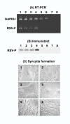
Figure 1
Figure 1
Presence of RSV in children with respiratory disease. (A) Detection of RSV-specific RNA. Collection of eye swabs and RT-PCR amplification of RSV F mRNA and "control" GAPDH mRNA segments were carried out as described under Methods. Only representative samples are shown. Lanes 1–4 = respiratory patients; 5,6 = healthy, uninfected individuals; 7,8 = same samples as 1 and 2, respectively, except that the RT reaction was not performed before PCR. (B) Presence of infectious RSV. Portions of the eye swab wash were added to HEp-2 monolayers to test for RSV growth as described under Methods. The lane numbers represent the same patient samples as in A. The P protein of RSV was detected by immunoblot; note that samples that contained viral RNA also contained infectious virus as judged by the intracellular synthesis of the P protein (lanes 1–4). (C) Syncytia in HEp-2 monolayers inoculated with samples #1 through 6 (the same numbers as in A and B). Note that samples that were RSV-positive (#1–4) in A and B formed syncytia, whereas RSV-negative samples (#5, 6) did not.
Figure 2
Figure 2
RSV growth in HCE cells. Growth was measured by (A) liberation of infectious virions and (B) synthesis of RSV P protein. At indicated times p.i., the released progeny virus was titered (closed circles in A) and the P protein was estimated by immunoblot (B) followed by densitometric analysis (open bars in A) as described under Methods.
Figure 3
Figure 3
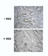
Syncytium in RSV-infected HCE cells. RSV infection of HCE monolayer was performed as described in Methods. At 72 h p.i. the monolayer was photographed under phase contrast [7]. Note that the infected cells are fused together (forming syncytia) in contrast to those in the mock-infected "control" monolayer.
Figure 4
Figure 4
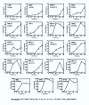
Induction of cytokine mRNA in RSV-infected HCE cells. RNA isolated from infected cells at different times p.i. were subjected to quantitative Real Time RT-PCR as described in Methods. The relative amounts of RNA were expressed as the ratio of mock-infected control value (fold induction). Each box represents a specific cytokine as named; the cognate receptor(s) are indicated in parenthesis. Each data point is derived from three independent infection experiments with the error bar as shown.
Figure 5
Figure 5
Quantitation of cytokines by antibody array. Immunoblot array of the indicated cytokines were performed and fold induction over mock-infected HCE cells were determined as described in Methods. IL-1α and IL-1β were subjected to greater scrutiny with closer time points because their mRNAs were not induced when tested in Fig. 4. Each bar is derived from three independent infection experiments with the error bar as shown. Note the strong elaboration of IL-1β, contrasting the little increase of IL-1α.
Figure 6
Figure 6
Activation of the HCE cell NF-κB by RSV. (A) Amounts of p65 and p50 subunits of NF-κB were determined by immunoblot of isolated nuclei. Note that a control transcription factor, Sp1, was not activated [6]. (B) Functional activation of NF-κB in RSV-infected cells was measured by luciferase induction from a reporter plasmid (pNFκB-Luc) as described in Methods. Where indicated, 10 or 20 mM NAC, Na-salicylate (SAL) or aspirin (ASP) was included in the reaction. Each bar is derived from three independent experiments with the standard error as indicated.

Similar articles

Cited by

References

    1. Thompson WW, Shay DK, Weintraub E, Brammer L, Cox N, Anderson LJ, Fukuda K. Mortality associated with influenza and respiratory syncytial virus in the United States. JAMA. 2003;289:179–186. doi: 10.1001/jama.289.2.179. - DOI - PubMed
    1. Maggon K, Barik S. New drugs and treatment for respiratory syncytial virus. Rev Med Virol. 2004;14:149–168. doi: 10.1002/rmv.423. - DOI - PubMed
    1. Marriott AC, Easton AJ. Reverse genetics of the Paramyxoviridae. Adv Virus Res. 1999;53:321–340. - PubMed
    1. Welliver RC. Respiratory syncytial virus and other respiratory viruses. Pediatr Infect Dis J. 2003;22:S6–S12. doi: 10.1097/00006454-200302001-00002. - DOI - PubMed
    1. Greenough A. Respiratory syncytial virus infection: clinical features, management, and prophylaxis. Curr Opin Pulm Med. 2002;8:214–217. doi: 10.1097/00063198-200205000-00011. - DOI - PubMed

Publication types

MeSH terms