Comparative Study
. 2004 Jul 15;18(14):1695-708.
doi: 10.1101/gad.1178604.

A signaling mucin at the head of the Cdc42- and MAPK-dependent filamentous growth pathway in yeast

Affiliations
Comparative Study

A signaling mucin at the head of the Cdc42- and MAPK-dependent filamentous growth pathway in yeast

Paul J Cullen et al. Genes Dev. .

Abstract

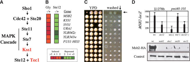
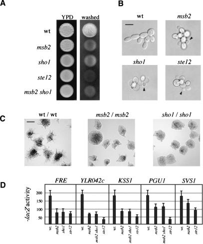
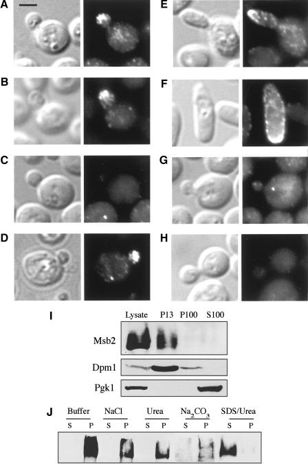
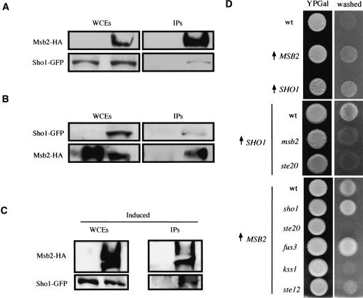
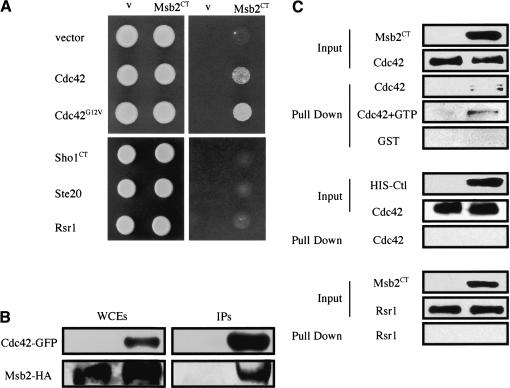
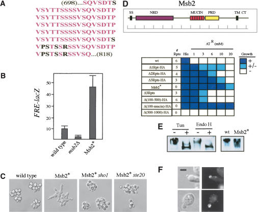
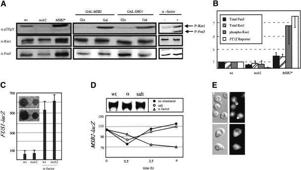
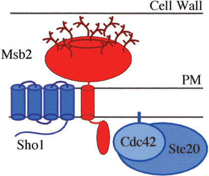
Signaling molecules such as Cdc42 and mitogen-activated protein kinases (MAPKs) can function in multiple pathways in the same cell. Here, we propose one mechanism by which such factors may be directed to function in a particular pathway such that a specific response is elicited. Using genomic approaches, we identify a new component of the Cdc42- and MAPK-dependent signaling pathway that regulates filamentous growth (FG) in yeast. This factor, called Msb2, is a FG-pathway-specific factor that promotes differential activation of the MAPK for the FG pathway, Kss1. Msb2 is localized to polarized sites on the cell surface and interacts with Cdc42 and with the osmosensor for the high osmolarity glycerol response (HOG) pathway, Sho1. Msb2 is glycosylated and is a member of the mucin family, proteins that in mammalian cells promote disease resistance and contribute to metastasis in cancer cells. Remarkably, loss of the mucin domain of Msb2 causes hyperactivity of the FG pathway, demonstrating an inhibitory role for mucin domains in MAPK pathway activation. Taken together, our data suggest that Msb2 is a signaling mucin that interacts with general components, such as Cdc42 and Sho1, to promote their function in the FG pathway.


Figures

Figure 1.
Figure 1.
MSB2 is an FG-pathway target. (A) The FG pathway with general factors (black) and FG-pathway-specific factors (red). (B) DNA microarray analysis. Targets of a glycosylation defect (left column, Gly-/+, pmi40-101 induced in YPD-/+ Man) that were also Ste12-dependent (right column, Ste12-/+, pmi40-101 ± ste12 induced in YPD-Man) are shown. (Red) Induced expression; (green) repressed expression (inset shows fold change). FUS1-HIS3 is a predicted target used as a control (Cullen et al. 2000). (C) Part of the genomic screen to isolate agar-invasion defective mutants. (Left panel) Colonies pinned to YPD for 4 d; (right panel) washed plate. Arrows point to the msb2 mutant. (D) MSB2 expression. (Upper panel) MSB2-lacZ expression. Cells of the indicated genotype and background containing pMSB2-lacZ were assayed for β-galactosidase activity. Wild-type (wt) or ste12 mutant ∑1278b cells were assayed over time for MSB2 expression; the 16-h time point is shown. Glycosylation mutant pmi40-101 containing or lacking the ste12 mutation (wild type [wt], pmi40-101, and ste12, pmi40-101 ste12) was incubated for 8 h ±Man as indicated. As shown, MSB2 expression in glycosylation mutants is partially induced by another factor. Numbers are in Miller Units, and error bars show standard deviation. (Lower two panels) Western blots. ∑1278b cells of the indicated genotypes expressing Msb2-HA were grown to mid-log phase in YPD, and extracts were assayed using antibodies against HA (upper panel) or a control protein (Dpm1, lower panel).
Figure 2.
Figure 2.
Msb2 is required for filamentous growth. (A) Plate-washing assay. Equal concentrations of cells of the indicated genotypes and ste4 were spotted onto YPD, incubated for 4 d (left), and washed (right). (B) Single-cell invasive-growth assay. Cells as in A were spread onto SC medium (lacking glucose), incubated for 16 h, and photographed at 100×. Arrows point to buds emerging from the proximal pole. Bar, 10 μm. (C) Pseudohyphal growth assay. Cells were spread onto low nitrogen (SLAHD) medium for 48 h and photographed at 20×. Bar, 50 μm. (D) Expression of FG-pathway reporters as indicated in given strains. Extracts prepared from cells in midlog phase SCD-LEU (SCD, synthetic complete dextrose) to select for reporter plasmids. All wild-type values were set at 180, and mutant values were adjusted by ratiometric division so that all values could be compared on the same scale. Numbers are in Miller Units.
Figure 3.
Figure 3.
Msb2 localization. (A-H) Immunolocalization of Msb2-HA. Treatments or genotypes are as follows: wild type (A,B); Latrunculin A (Lat A; C); DMSO (D); GAL-MSB2-HA (E,F); ste12 (G); no tag (H). (Left panels) DIC; (right panels) FITC. Bar, 5 μm. (I,J) Subcellular localization of Msb2. (I) Cells expressing Msb2-HA were solubilized and cell extracts were separated by centrifugation. Lysate, supernatnat (S), and pellet (P) fractions are shown ([13] 13,000XG; [100] 100,000XG). Western blotting was performed using antibodies against HA, Dpm1 (an ER integral-membrane protein), and Pgk1 (a soluble cytoplasmic protein) as indicated. (J) P13 fraction analysis. Treatments were lysis buffer alone (Buffer) or with NaCl (0.5 M), urea (5 M), sodium bicarbonate (100 mM Na2CO3 at pH 11), or SDS (5%) and urea (8 M).
Figure 4.
Figure 4.
Msb2 and Sho1 interact and function together in the FG pathway. (A) Immunoprecipitation of Msb2-HA also precipitates Sho1. Western blots of whole-cell extracts (WCEs) and immunoprecipitations (IPs) are shown, probed using antibodies specific for HA (Msb2-HA) and GFP (Sho1-GFP). (B) Immunoprecipitation of Sho1-GFP also precipitates Msb2-HA. Labels are as in A. (C) Sho1-GFP is coprecipitated at higher concentrations by Msb2-HA in cells undergoing FG. Induced refers to cells undergoing FG from which extracts for coIPs were prepared; see Materials and Methods for details. (Msb2 is more abundant under this condition, presumably because of to FG-pathway induction of MSB2 expression. However, the amount of IPed Msb2 is similar under induced and uninduced conditions; under inducing conditions, more Sho1 is precipitated. (D) Cells overproducing Msb2 or Sho1 in FG-pathway mutant backgrounds. Cells were spotted onto YP + 2% galactose (YPGal; left panels; [↑] GAL1 promoter) and washed after 48 h (right panels).
Figure 5.
Figure 5.
Msb2 interacts with Cdc42. (A) Two-hybrid analysis. GBD-Msb2CT in strains containing GAD fusions to GTPases Cdc42 and Rsr1 and FG-pathway components as shown. Growth was scored on SCD-URA-LEU-HIS + 4 mM aminotriazole (AT). (v) Vector. (B) Msb2-HA and Cdc42-GFP interact by coIP analysis. IP of Cdc42-GFP in IP buffer with 1% NP-40 using antibodies specific for GFP (upper panels) coprecipitates Msb2-HA (lower panels). The WCE and IPs are shown. (C) Cdc42 and Msb2CT interact in vitro. Fusion proteins were expressed and purified from E. coli and incubated together in binding buffer containing BSA (Input) and GTP as indicated. After a 15-min incubation, Co+ beads that recognize the HIS epitope on the Msb2CT and Ctl [Urm1] fusions were added to the reaction for 15 min, and HIS-tagged proteins were isolated by low-speed centrifugation (Pull Down). Western blotting was used to detect the GST epitopes on the Cdc42, GST, and Rsr1 fusions, and the HIS epitopes on the Msb2CT and Ctl [Urm1] fusions. The inputs for Cdc42 + GTP and GST reactions had equivalent input protein amounts to that shown for the inputs in the top two panels (data not shown).
Figure 6.
Figure 6.
Properties of the Msb2 mucin. (A) The mucin domain of Msb2. Identical amino acids (pink) and nonidentical amino acids (black) are shown. (B) FRE-lacZ expression in strains lacking Msb2 or containing Msb2*. Numbers are in Miller Units. (C) Morphological phenotypes associated with Msb2*. Cells were grown to saturation in YEPD medium and examined at 100×. (D) Hyperactive variants of the Msb2 protein. (Inset) The Msb2 protein. Shown are the N-terminal signal sequence (SS), the N-terminal negative regulatory domain (NRD, purple), the mucin repeats (MUCIN, pink), the positive regulatory domain (PRD, yellow), the transmembrane domain (TM), and the cytoplasmic tail (CT). Bar in 100-amino acid increments. Strains with indicated alleles of Msb2 were tested for expression of the FUS1-HIS3 reporter by growth on SCD-His + aminotriazole (AT) at the concentrations shown. Growth was scored over separate trials: (+) growth, dark blue; (+/-) spotty growth, light blue; (-) no growth, white. (E) Msb2 is glycosylated. (Left panel) Cells containing Msb2-HA were grown to mid-log phase in YPD and treated with DMSO (lane 1) or tunicamycin in DMSO (lane 2) for 2 h, and extracts were prepared for SDS-PAGE and Western analysis using anti-HA antibodies. (Lanes 3,4) Endo H treatment (right two lanes) was performed on cell extracts derived from mid-log-phase cells. (Right panel) Msb2-HA (wt) or Msb2* cells were grown to mid-log phase, and extracts were prepared and analyzed as above. (F) Localization of Msb2-HA in cells treated with tunicamycin (upper two panels). Localization of Msb2* (lower two panels). Bar, 5 μm. (Left panels) DIC; (right panels) FITC. For wild-type reference, see Figure 3A.
Figure 7.
Figure 7.
Msb2 is an FG-pathway-specific factor. (A) Western blot of P∼Kss1 (upper band, upper panel), P∼Fus3 (lower band, upper panel), total Kss1 (middle panel), and total Fus3 (bottom panel) from strains containing the indicated genotypes and ste4. (B) Total Fus3 protein, total and dually phosphorylated Kss1 protein, and FRE-lacZ from YEpU-FT1Z were quantified in two independent experiments and normalized to wild type. (C) FUS1-lacZ expression and halo assays. Wild-type (wt) or msb2 cells were incubated in ±30 μM α-factor for 4 h and tested for FUS1-lacZ expression. Numbers are in Miller Units. (Inset) Halo assay. Cells were spread onto YPD plates to which 2 or 10 μL of 1 μg/μL α-factor was applied. (D) Wild-type cells containing MSB2-lacZ in mid-log phase were incubated ±0.9 M NaCl or ±30 μM α-factor. Numbers are in Miller Units. (Upper panel) Western blot of Msb2-HA from same cells treated with salt or α-factor: (α) α-factor. (E) Msb2-HA localization in cells treated with 30 μM α-factor for 3 h (lower panels) or untreated (upper panels).
Figure 8.
Figure 8.
Model for the protein complex at the head of the FG pathway. Msb2 (in red) is composed of a large extracellular domain, which contains the mucin repeats (bright red oval) and is glycosylated (as shown), and integral-membrane and cytoplasmic domains. General factors Sho1, Cdc42 (isoprenylated and attached to the plasma membrane; PM), and Ste20 all shown in blue are recruited to the FG pathway by Msb2. The interaction between Cdc42 and the cytoplasmic domain of Msb2 is indicated, as is the interaction between Msb2 and Sho1, which is stimulated by FG-pathway activation. An interaction between the cytoplasmic domains of the Msb2 and Sho1 proteins was not detected; thus, they may interact in their membrane-spanning or extracellular domains.

References

    1. Agrawal B., Gendler, S.J., and Longenecker, B.M. 1998a. The biological role of mucins in cellular interactions and immune regulation: Prospects for cancer immunotherapy. Mol. Med. Today 4: 397-403. - PubMed
    1. Agrawal B., Krantz, M.J., Parker, J., and Longenecker, B.M. 1998b. Expression of MUC1 mucin on activated human T cells: Implications for a role of MUC1 in normal immune regulation. Cancer Res. 58: 4079-4081. - PubMed
    1. Baldi P. and Long, A.D. 2001. A Bayesian framework for the analysis of microarray expression data: Regularized t-test and statistical inferences of gene changes. Bioinformatics 17: 509-519. - PubMed
    1. Bardwell L., Cook, J.G., Voora, D., Baggott, D.M., Martinez, A.R., and Thorner, J. 1998a. Repression of yeast Ste12 transcription factor by direct binding of unphosphorylated Kss1 MAPK and its regulation by the Ste7 MEK. Genes & Dev. 12: 2887-2898. - PMC - PubMed
    1. Bardwell L., Cook, J.G., Zhu-Shimoni, J.X., Voora, D., and Thorner, J. 1998b. Differential regulation of transcription: Repression by unactivated mitogen-activated protein kinase Kss1 requires the Dig1 and Dig2 proteins. Proc. Natl. Acad. Sci. 95: 15400-15405. - PMC - PubMed

Publication types

MeSH terms

Substances