Comparative Study
. 2004 Jul 15;18(14):1737-52.
doi: 10.1101/gad.294904.

Tal, a Tsg101-specific E3 ubiquitin ligase, regulates receptor endocytosis and retrovirus budding

Affiliations
Comparative Study

Tal, a Tsg101-specific E3 ubiquitin ligase, regulates receptor endocytosis and retrovirus budding

Ido Amit et al. Genes Dev. .

Abstract

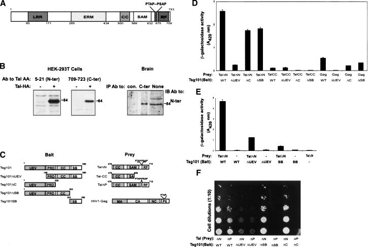
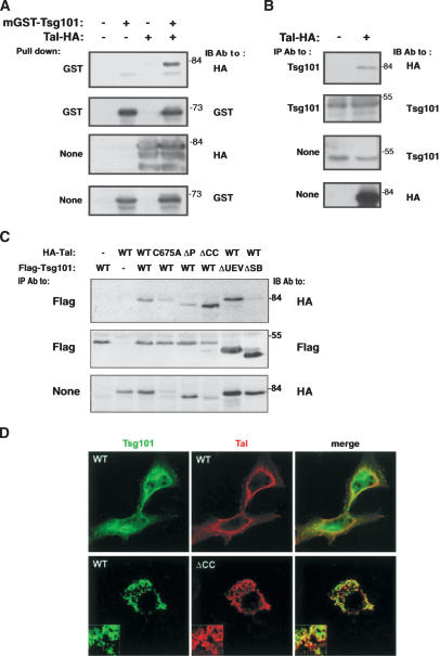
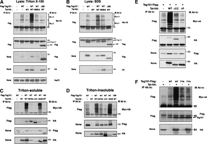
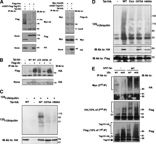
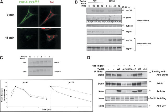
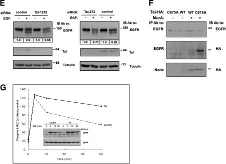
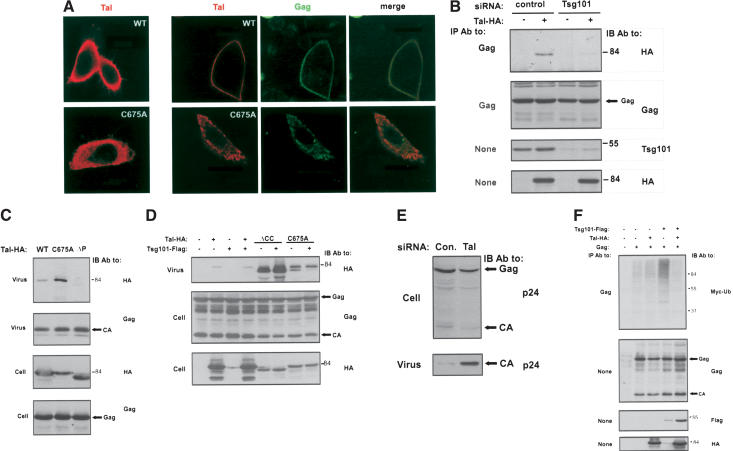
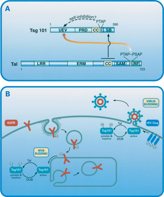
The tumor suppressor gene 101 (tsg101) regulates vesicular trafficking processes in yeast and mammals. We report a novel protein, Tal (Tsg101-associated ligase), whose RING finger is necessary for multiple monoubiquitylation of Tsg101. Bivalent binding of Tsg101 to a tandem tetrapeptide motif (PTAP) and to a central region of Tal is essential for Tal-mediated ubiquitylation of Tsg101. By studying endocytosis of the epidermal growth factor receptor and egress of the human immunodeficiency virus, we conclude that Tal regulates a Tsg101-associated complex responsible for the sorting of cargo into cytoplasm-containing vesicles that bud at the multivesicular body and at the plasma membrane.


Figures

Figure 1.
Figure 1.
Domain structure of human Tal and binding with Tsg101 in yeast. (A) Schematic diagram of human Tal, showing the approximate boundaries of the leucine-rich repeats (LRRs), ezrin-radixin-moesin (ERM) domain, coiled-coil (CC) region, α sterile alpha motif (SAM), a RING finger (RF), and a double PTAP motif. (B) HA-tagged hTal was immunoprecipitated (IP) from extracts of HEK-293T cells transfected with either the respective plasmid, or an empty vector (-). The rabbit antibodies used for IP are specific to peptides corresponding to the indicated amino acids (AA) of hTal. An antibody to HA was used for immunoblotting (IB). Alternatively, extracts of mouse brain were subjected to IB, either directly (None) or after IP. (C) Schematic diagrams of Tsg101, Tal, and Gag proteins, along with their mutants. The following domains of Tsg101 are indicated: ubiquitin E2 variant (UEV) region, proline-rich domain (PRD), coiled coil (CC), and steadiness box (SB). The PTAP motif of Gag is indicated inside the p6 domain. Other Gag domains are matrix (MA), capsid (CA), and nucleocapsid (NC). (D,E) The interaction between Tal (or a LexA control protein; -) and Tsg101, either wild-type (WT) or the indicated mutants, was determined in yeast by using reporter gene activation. The histogram represents the average (±S.D.) activity of β-galactosidase measured in three separate assays using O-nitrophenyl-β-D-galactopyranoside as a substrate. (F) Yeast cells transformed by the indicated pairs of plasmids were subjected to serial dilutions and plated on selective (Trp-Leu-His minus) medium.
Figure 2.
Figure 2.
Bimodal interaction and partial colocalization of Tal and Tsg101. (A) Extracts of HEK-293T cells coexpressing mGST-Tsg101 and HA-hTal were subjected to a pull-down assay using glutathione-agarose beads and IB, as indicated. (B) Extracts of HEK-293T cells transiently expressing HA-hTal were subjected to IP with an antibody directed to the endogenous Tsg101 protein. Cell lysates and IPs were analyzed as indicated. (C) HEK-293T cells coexpressing various forms of HA-hTal and Flag-Tsg101 were analyzed for coprecipitation. (D) HeLa cells expressing HA-hTal (either wild type [WT] or ΔCC) and Flag-Tsg101 were fixed, permeabilized, and stained with primary and fluorescently labeled secondary antibodies, prior to confocal microscopy.
Figure 3.
Figure 3.
Tal increases ubiquitylation of Tsg101 and affects its solubility in a RING- and PTAP-dependent manner. (A,B) HEK-293T cells were cotransfected with plasmids encoding Flag-Tsg101 (either wild type [WT] or ΔSB), HA-Tal (either wild type [WT] or H695A), and Myc-ubiquitin. Forty-eight hours after transfection, cells were divided into two portions: The first (75% of cell population) was extracted by using Triton X-100 (A), and the remainder was extracted in SDS (B). IB of immunoprecipitates or whole extracts was performed with the indicated antibodies. Oligo- and multiubiquitylated forms of Tsg101 are labeled. (C,D) HEK-293T cells transiently expressing the indicated proteins, along with Myc-Ub, were sequentially extracted in Triton X-100 (C) and SDS (D) and analyzed as indicated. (E,F) HEK-293T cells transiently expressing the indicated proteins, along with Myc-Ub, were extracted in Triton X-100 and analyzed as indicated. The TYN mutant of Tsg101 represents a protein whose TYN motif of the UEV domain (residues 67-69) has been replaced with a triplet of alanines.
Figure 4.
Figure 4.
Tal monoubiquitylates Tsg101 and undergoes self-ubiquitylation. (A) HEK-293T cells transiently expressing combinations of Hrs or Vps28, along with Tal, Tsg101, and either Myc- (left) or Flag- (right) tagged ubiquitin, were extracted in Triton X-100 and analyzed. (B) The state of ubiquitylation of WT-hTal or its indicated mutants was examined by coexpressing a Flag-tagged ubiquitin in HEK-293T cells. (C) The indicated forms of HA-Tal were immunoprecipitated from extracts of HEK-293T cells and incubated with recombinant E1, E2 (Ubc-H5B), and 125I-labeled ubiquitin. Ubiquitylated products were resolved by gel electrophoresis and detected by autoradiography. (D) An immunoprecipitated Flag-Tsg101 was incubated with extracts of HEK-293T cells transfected with either a control plasmid (Con), or vectors encoding the indicated forms of hTal. After extensive washing, the Tsg101-Tal complex was subjected to ubiquitylation in vitro using a radiolabeled ubiquitin. A control reaction was performed in the absence of cell extract (lane labeled -). (E) Extracts of HEK-293T cells expressing Flag-Tsg101, HA-, and Myc-tagged ubiquitin (either wild type [WT] or 4KR), and either GFP-Tal or a control plasmid, were subjected to immunoprecipitation (first IP) with an anti-Flag antibody. A portion (10%) of the beads was directly analyzed and the rest eluted at 95°C and subjected to IP with an anti-HA antibody (second IP). Likewise, a portion (10%) of the beads was directly analyzed and the rest eluted, subjected to IP with an anti-Myc antibody (third IP), and analyzed by electrophoresis. A closed arrowhead indicates the location of nonubiquitylated Tsg101, and an open arrowhead marks the monoubiquitylated form.
Figure 5.
Figure 5.
A catalytically inactive mutant of hTal stably associates with EGFRs and accelerates their endocytic degradation. (A) HeLa cells expressing HA-hTal were preincubated at 4°C with EGF conjugated to AlexaFluor 488. Thereafter, cells were incubated for 15 min at 37°C, permeabilized, and stained with anti-HA antibodies, followed by a fluorescent secondary antibody. (B) HEK-293 cells stably expressing the ecdysone receptor were transfected with plasmids that express HA-hTal (wild type [WT] or C675A) under the control of an ecdysone-inducible promoter. The indicated stable clones were incubated without or with Muristerone A (2 μM) for the indicated time intervals and thereafter cells were extracted in Triton X-100. The insoluble material was extracted in SDS, and both fractions were directly analyzed. Numbers below lanes indicate quantification of signals normalized to the respective tubulin signal. (C) Chinese hamster ovary (CHO) cells transiently transfected with an EGFR plasmid, along with either a C675A-Tal (solid line) or an empty vector (Control; broken line), were preincubated in cysteine- and methionine-free medium prior to a 20-min-long pulse of [35S]-labeled amino acids. Thereafter, cells were chased at 37°C in fresh medium for the indicated time intervals. An autoradiogram of the immunoprecipitated EGFR is shown, along with the respective quantification of the precursor (p140) and mature (p170) forms of EGFR. (D) CHO cells transiently coexpressing EGFR, Flag-Tsg101, and the indicated forms of HA-hTal were surface biotinylated 48 h after transfection. A GFP expression vector was used to monitor transfection efficiency. Cell extracts were analyzed as indicated. (E) HeLa-SS6 cells were transfected with the indicated siRNA oligonucleotides (50 nM each). Forty-eight hours posttransfection, cells were starved for 6 h in the absence of serum, and then stimulated with EGF (20 ng/mL) for 1 h. Whole-cell extracts were immunoblotted with the indicated antibodies. The EGFR signals were quantified and normalized to the respective levels of tubulin. (F) HEK-293 cells expressing HA-hTal (wild type [WT] or C675A) from a Muristerone-inducible promoter were incubated without or with Muristerone A (2 μM) for 48 h. Cell extracts were tested for coimmunoprecipitation of hTal and EGFR. (G) HEK-293T cells were cotransfected with a GFP-ERK2 plasmid and either a vector encoding for HA-hTal or a control empty plasmid. Thirty-six hours posttransfection, cells were starved for 8 h in the absence of serum, and then stimulated with EGF (100 ng/mL) for the indicated time intervals. Whole-cell extracts were immunoblotted with antibodies to the doubly phosphorylated ERK (pERK) or a general ERK antibody (gERK). Shown are the resulting immunoblots (inset) and quantification of the active ERK signal.
Figure 5.
Figure 5.
A catalytically inactive mutant of hTal stably associates with EGFRs and accelerates their endocytic degradation. (A) HeLa cells expressing HA-hTal were preincubated at 4°C with EGF conjugated to AlexaFluor 488. Thereafter, cells were incubated for 15 min at 37°C, permeabilized, and stained with anti-HA antibodies, followed by a fluorescent secondary antibody. (B) HEK-293 cells stably expressing the ecdysone receptor were transfected with plasmids that express HA-hTal (wild type [WT] or C675A) under the control of an ecdysone-inducible promoter. The indicated stable clones were incubated without or with Muristerone A (2 μM) for the indicated time intervals and thereafter cells were extracted in Triton X-100. The insoluble material was extracted in SDS, and both fractions were directly analyzed. Numbers below lanes indicate quantification of signals normalized to the respective tubulin signal. (C) Chinese hamster ovary (CHO) cells transiently transfected with an EGFR plasmid, along with either a C675A-Tal (solid line) or an empty vector (Control; broken line), were preincubated in cysteine- and methionine-free medium prior to a 20-min-long pulse of [35S]-labeled amino acids. Thereafter, cells were chased at 37°C in fresh medium for the indicated time intervals. An autoradiogram of the immunoprecipitated EGFR is shown, along with the respective quantification of the precursor (p140) and mature (p170) forms of EGFR. (D) CHO cells transiently coexpressing EGFR, Flag-Tsg101, and the indicated forms of HA-hTal were surface biotinylated 48 h after transfection. A GFP expression vector was used to monitor transfection efficiency. Cell extracts were analyzed as indicated. (E) HeLa-SS6 cells were transfected with the indicated siRNA oligonucleotides (50 nM each). Forty-eight hours posttransfection, cells were starved for 6 h in the absence of serum, and then stimulated with EGF (20 ng/mL) for 1 h. Whole-cell extracts were immunoblotted with the indicated antibodies. The EGFR signals were quantified and normalized to the respective levels of tubulin. (F) HEK-293 cells expressing HA-hTal (wild type [WT] or C675A) from a Muristerone-inducible promoter were incubated without or with Muristerone A (2 μM) for 48 h. Cell extracts were tested for coimmunoprecipitation of hTal and EGFR. (G) HEK-293T cells were cotransfected with a GFP-ERK2 plasmid and either a vector encoding for HA-hTal or a control empty plasmid. Thirty-six hours posttransfection, cells were starved for 8 h in the absence of serum, and then stimulated with EGF (100 ng/mL) for the indicated time intervals. Whole-cell extracts were immunoblotted with antibodies to the doubly phosphorylated ERK (pERK) or a general ERK antibody (gERK). Shown are the resulting immunoblots (inset) and quantification of the active ERK signal.
Figure 6.
Figure 6.
Tal interacts with Tsg101 and Gag along the exocytic pathway of HIV-1. (A) Cultures of HeLa-SS6 cells expressing either wild type (WT) or C675A-hTal were treated with either a control vector (left column), or a Gag-GFP-encoding plasmid. Fixation, staining, and confocal visualization were performed 6 h later. (B) HeLa-SS6 cells were transfected with the indicated siRNA oligonucleotides, and 24 h later a second transfection was performed with vectors encoding HIV-1 Gag (pNLenv-1; Schubert et al. 1995) and HA-hTal (or a control plasmid). Cells were extracted 24 h later, and coimmunoprecipitation of Tal and Gag was tested by using the respective antibodies. (C,D) HEK-293T cells were cotransfected with the pNLenv-1 vector encoding HIV-1 Gag, along with the indicated Tal and Tsg101 plasmids. Cells and VLPs were analyzed 48 h posttransfection (CA; capsid). (E) HeLa-SS6 cells were transfected with a Tal-specific siRNA, which starts at nucleotide 1252, and a control inverted sequence (50 nM each). Forty-eight hours later, cells were cotransfected with the indicated oligonucleotides (25 nM) along with pNLenv-1 (1 μg). The presence of Gag in VLPs or in cytoplasmic extracts was analyzed 24 h later. (F) HEK-293T cells were cotransfected with pNLenv-1, along with a Myc-ubiquitin vector and the indicated Flag-Tsg101 and HA-hTal plasmids. Forty-eight hours posttransfection, cells were extracted and analyses performed either directly or after IP with anti-Gag antibodies.
Figure 7.
Figure 7.
Structural and functional interactions between Tal and Tsg101. (A) The domain structures of Tal and Tsg101 are depicted, along with their intermolecular binding specificities (for abbreviations, see Fig. 1). Note that the UEV domain of Tsg101 binds the double PTAP motif of Tal, and a distinct site binds a monomeric ubiquitin (not shown). Secondary interactions between Tal and Tsg101 involve a region encompassing the coiled-coil (CC) domain of Tal and the steadiness box (SB) of Tsg101. Potentially, both binding sites of the UEV domain may be blocked intramolecularly through binding to the C-terminally located PTAP motif and to a monomeric ubiquitin conjugated by Tal. (B) Model illustrating the role of the Tal-Tsg101 complex in budding of vesicles into the lumen of the multivesicular body (MVB) and in virus budding. Accordingly, Tsg101 sorts cargo proteins like the epidermal growth factor receptor (EGFR) and HIV Gag into budding structures. Tal-mediated ubiquitylation of Tsg101 inactivates this sorting function and concomitantly translocates Tsg101 from relatively insoluble membrane subdomains. Presumably, the coordinated action of Tal and a deubiquitylation enzyme (DUB) enables recycling of Tsg101 and reloading of cargo.

Similar articles

Cited by

References

    1. Babst M., Odorizzi, G., Estepa, E.J., and Emr, S.D. 2000. Mammalian tumor susceptibility gene 101 (TSG101) and the yeast homologue, Vps23p, both function in late endosomal trafficking. Traffic 1: 248-258. - PubMed
    1. Bache K.G., Brech, A., Mehlum, A., and Stenmark, H. 2003a. Hrs regulates multivesicular body formation via ESCRT recruitment to endosomes. J. Cell Biol. 162: 435-442. - PMC - PubMed
    1. Bache K.G., Raiborg, C., Mehlum, A., and Stenmark, H. 2003b. STAM and Hrs are subunits of a multivalent ubiquitin-binding complex on early endosomes. J. Biol. Chem. 278: 12513-12521. - PubMed
    1. Bishop N. and Woodman, P. 2001. TSG101/mammalian VPS23 and mammalian VPS28 interact directly and are recruited to VPS4-induced endosomes. J. Biol. Chem. 276: 11735-11742. - PubMed
    1. Cadavid A.L., Ginzel, A., and Fischer, J.A. 2000. The function of the Drosophila fat facets deubiquitinating enzyme in limiting photoreceptor cell number is intimately associated with endocytosis. Development 127: 1727-1736. - PubMed

Publication types

MeSH terms