

Enhanced expression of transforming growth factor beta 1 in the rat brain after a localized cerebral injury
- PMID: 1525658
- PMCID: PMC4310563
- DOI: 10.1016/0006-8993(92)91000-5
Enhanced expression of transforming growth factor beta 1 in the rat brain after a localized cerebral injury
Abstract
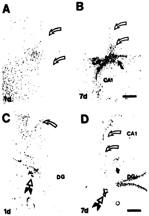
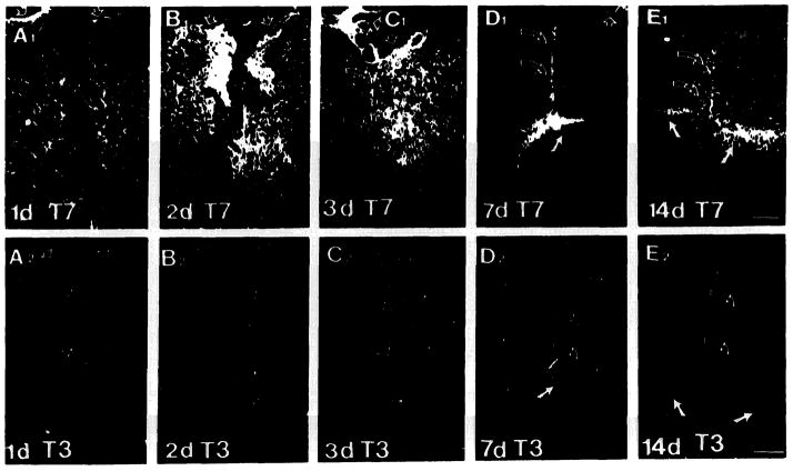
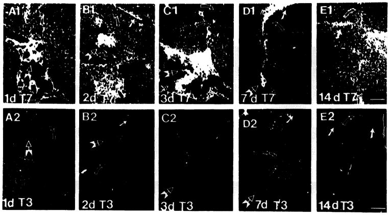
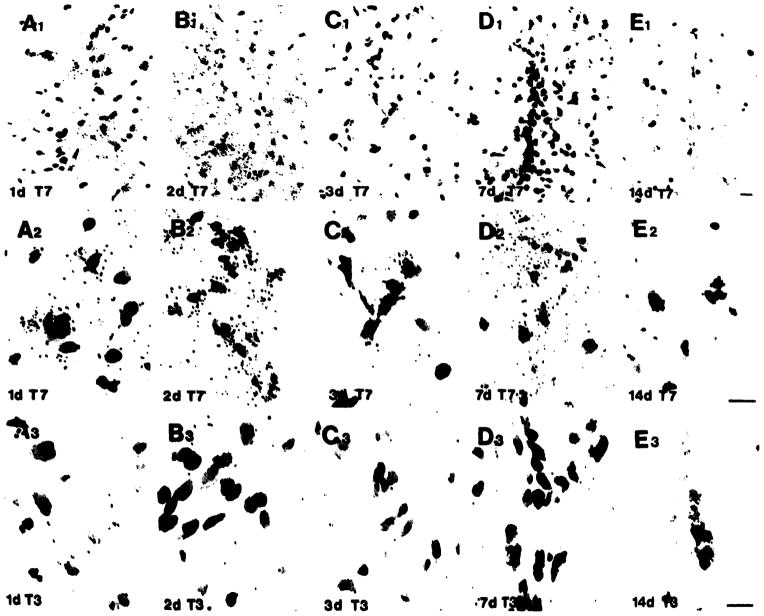
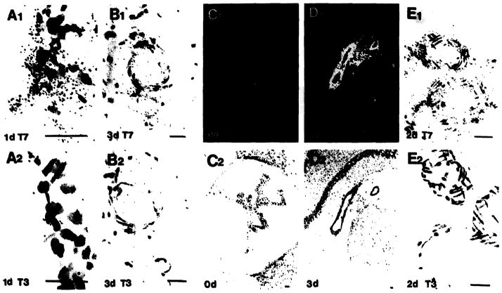
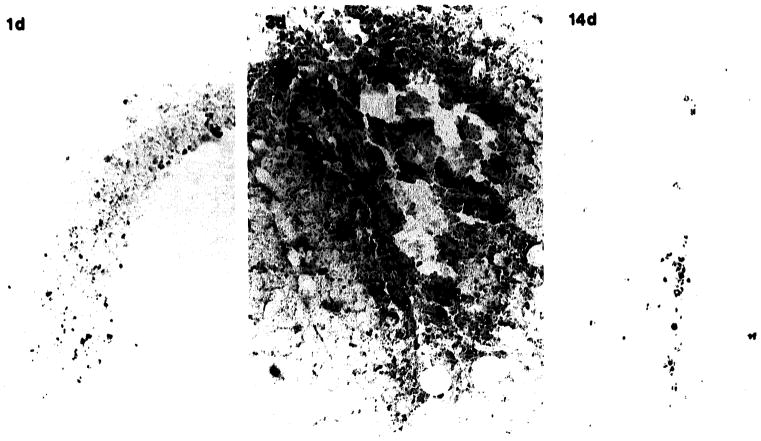
It is becoming clear that transforming growth factor beta (TGF beta) may be a key factor regulating inflammatory and tissue specific wound responses. Because the formation of a glial-collagen scar at CNS lesion sites is thought to contribute to the pathology associated with penetrating CNS injuries, and because in the periphery TGF beta 1 stimulates fibroblast deposition of scar tissue, we used in situ hybridization and immunohistochemistry to investigate the effect of a defined cerebral lesion on the local expression of TGF beta 1. Induction of TGF beta 1 mRNA and protein is relatively diffuse in the neuropile around the margins of the lesion at 1, 2 and 3 days, but becomes localized to the region of the glial scar at 7 and 14 days. The signal intensity for TGF beta 1 mRNA and protein is maximal between 2 and 3 days and decreases between 7 and 14 days after lesion. The predominant cell types in the neuropile localizing TGF beta 1 mRNA and protein have the morphological characteristics of astrocytes, although macrophages are also detected. An induction of TGF beta 1 mRNA was also observed in endothelial cells of the meninges, hippocampal fissure and choroid plexus, at 2 and 3 days. However, this is dramatically reduced by 7 days and has disappeared by 14 days. These results suggest a role for TGF beta 1, not only in inflammation, but also in the tissue-specific glial scar formation that occurs in the CNS. Furthermore, they suggest a potential therapeutic use of TGF beta 1 antagonists in the CNS to help limit the pathogenesis associated with matrix deposition in the wound.
Figures

Similar articles
-
Transforming growth factor beta 1 and fibronectin messenger RNA in rat brain: responses to injury and cell-type localization.Neuroscience. 1993 Jun;54(4):893-907. doi: 10.1016/0306-4522(93)90583-2. Neuroscience. 1993. PMID: 8341423
-
Transforming growth factor-beta 1 in the rat brain: increase after injury and inhibition of astrocyte proliferation.J Cell Biol. 1992 Apr;117(2):395-400. doi: 10.1083/jcb.117.2.395. J Cell Biol. 1992. PMID: 1560032 Free PMC article.
-
Effects of transforming growth factor beta 1 on scar production in the injured central nervous system of the rat.Eur J Neurosci. 1994 Mar 1;6(3):355-63. doi: 10.1111/j.1460-9568.1994.tb00278.x. Eur J Neurosci. 1994. PMID: 8019673
-
The role of the fetal fibroblast and transforming growth factor-beta in a model of human fetal wound repair.Semin Pediatr Surg. 1996 Aug;5(3):165-74. Semin Pediatr Surg. 1996. PMID: 8858763
-
Smad3 null mice display more rapid wound closure and reduced scar formation after a stab wound to the cerebral cortex.Exp Neurol. 2007 Jan;203(1):168-84. doi: 10.1016/j.expneurol.2006.08.006. Epub 2006 Sep 22. Exp Neurol. 2007. PMID: 16996058
Cited by
-
TGF-beta superfamily members promote survival of midbrain dopaminergic neurons and protect them against MPP+ toxicity.EMBO J. 1995 Feb 15;14(4):736-42. doi: 10.1002/j.1460-2075.1995.tb07052.x. EMBO J. 1995. PMID: 7882977 Free PMC article.
-
Chondroitin sulfate proteoglycan and tenascin in the wounded adult mouse neostriatum in vitro: dopamine neuron attachment and process outgrowth.J Neurosci. 1996 Dec 15;16(24):8005-18. doi: 10.1523/JNEUROSCI.16-24-08005.1996. J Neurosci. 1996. PMID: 8987827 Free PMC article.
-
Mechanisms of hydrocephalus after intraventricular haemorrhage in adults.Stroke Vasc Neurol. 2016 Feb 16;1(1):23-27. doi: 10.1136/svn-2015-000003. eCollection 2016 Mar. Stroke Vasc Neurol. 2016. PMID: 28959460 Free PMC article. Review.
-
TGF-beta enhances effector Th1 cell activation but promotes self-regulation via IL-10.J Immunol. 2010 May 15;184(10):5628-36. doi: 10.4049/jimmunol.1000288. Epub 2010 Apr 14. J Immunol. 2010. PMID: 20393141 Free PMC article.
-
Homeostatic capabilities of the choroid plexus epithelium in Alzheimer's disease.Cerebrospinal Fluid Res. 2004 Dec 10;1(1):3. doi: 10.1186/1743-8454-1-3. Cerebrospinal Fluid Res. 2004. PMID: 15679944 Free PMC article.
References
-
- Anderson KJ, Dam D, Lee S, Cotman CW. Basic fibroblast growth factor prevents death of lesioned cholinergic neurons in vivo. Nature. 1988;332:360–361. - PubMed
-
- Assoian RK, Komoriya A, Myers CA, Miller DM, Sporn MB. Transforming growth factor-β in human platelets. J Biol Chem. 1983;258:7155–7160. - PubMed
-
- Beck DW, Hart MN, Cancilla PA. The role of the macrophage in microvascular regeneration following brain injury. J Neuropathol Exp Neurol. 1983;42:601–614. - PubMed
-
- Bensaid M, Malecaze F, Bayard F, Tauber JP. Opposing effects of basic fibroblast growth factor and transforming growth factor-β on the proliferation of cultured bovine retinal capillary endothelial (BREC) cells. Exp Eye Res. 1989;48:791–799. - PubMed
-
- Berry M, Maxwell WL, Logan A, Mathewson A, McConnell P, Ashhurst DE, Thomas GH. Deposition of scar tissue in the central nervous system. Acta Neurochir Suppl. 1983;32:31–35. - PubMed
Publication types
MeSH terms
Substances
Grants and funding
LinkOut - more resources
Full Text Sources
Other Literature Sources
