. 2004 Aug 18;23(16):3314-24.
doi: 10.1038/sj.emboj.7600316. Epub 2004 Jul 15.

Nucleosomes containing the histone variant H2A.Bbd organize only 118 base pairs of DNA

Affiliations

Nucleosomes containing the histone variant H2A.Bbd organize only 118 base pairs of DNA

Yunhe Bao et al. EMBO J. .

Abstract

H2A.Bbd is an unusual histone variant whose sequence is only 48% conserved compared to major H2A. The major sequence differences are in the docking domain that tethers the H2A-H2B dimer to the (H3-H4)(2) tetramer; in addition, the C-terminal tail is absent in H2A.Bbd. We assembled nucleosomes in which H2A is replaced by H2A.Bbd (Bbd-NCP), and found that Bbd-NCP had a more relaxed structure in which only 118+/-2 bp of DNA is protected against digestion with micrococcal nuclease. The absence of fluorescence resonance energy transfer between the ends of the DNA in Bbd-NCP indicates that the distance between the DNA ends is increased significantly. The Bbd docking domain is largely responsible for this behavior, as shown by domain-swap experiments. Bbd-containing nucleosomal arrays repress transcription from a natural promoter, and this repression can be alleviated by transcriptional activators Tax and CREB. The structural properties of Bbd-NCP described here have important implications for the in vivo function of this histone variant and are consistent with its proposed role in transcriptionally active chromatin.


Figures
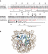
Figure 1
Figure 1
Unique features of H2A.Bbd. (A) Sequence alignment of mouse H2A.1 (gi: 121961), human H2A.1 (gi: 106265), human H2A.Bbd (gi: 15553137) and X. laevis H2A (gi: 121966). Intervals of 10 amino acids for H2A.1 (filled circles) and H2A.Bbd (open circles) are indicated. Sequence differences are shown in red. The docking domain is indicated by a dashed line. Secondary structure elements of the histone fold (α1, α2 and α3) and extensions (αN and αC) are indicated. Truncated and domain-swapped regions are indicated by arrows. (B) Structure of Xla-NCP to indicate the structural context of major sequence differences in H2A.Bbd. H2A is shown in yellow, H2B in red, H3 in blue and H4 in green. The H2A docking domain is colored orange and the H2A C-terminus (H2A C) is indicated. The black arrow indicates the approximate C-terminus of H2A.Bbd, based on the sequence alignment shown in (A). Sites of truncation and domain swap are indicated by a star.
Figure 2
Figure 2
H2A.Bbd does not refold to a histone octamer with H2B, H3 and H4. (A) Gel filtration of refolding reactions from mouse histones (octamer, solid line) and mouse H3, H4, H2B and H2A.Bbd (tetramer and dimer, dashed line). (B) Fractions containing mouse tetramer (mmT, lane 2), H2A.Bbd–H2B dimer (BD, lane 1) and mouse octamer (mmO, lane 3) were analyzed by 18% SDS–PAGE, stained with Coomassie brilliant blue.
Figure 3
Figure 3
Bbd-NCP is structurally altered. (A) Salt gradient reconstituted Xla-NCP (lanes 1 and 2), Bbd-NCP (lanes 3 and 4) and mm-NCP (lanes 5 and 6), before (−) and after (+) a 1 h incubation at 37°C, were analyzed by 5% native PAGE and stained with Coomassie blue. (B) Analysis of the histone content of the two Bbd-NCP nucleosomal bands. The upper band (Ub; lane 5) and lower band (Lb; lane 2) of Bbd-NCP were excised from the native gel and analyzed by 18% SDS–PAGE. H2A.Bbd–H2B dimer: (H3–H4)2 tetramer mixtures of 2:1 (Oct, lane 4) and 1:1 (Hex, lane 3) were used as controls. (C) yNAP-1-reconstituted mm-NCP (lanes 1 and 2) and Bbd-NCP (lanes 5 and 6) were compared with salt gradient reconstituted NCPs (lanes 3 and 4, respectively) on a 5% native gel, stained with ethidium bromide. (D) Stability of mm-NCP and Bbd-NCP at elevated ionic strength. Heat-shifted NCPs (lanes 2 and 7) were incubated at 37°C for 1 h in the presence of 200, 400 and 600 mM KCl (lanes 3, 4, 5 and 8, 9, 10). (E) The integral distributions of sedimentation coefficients, G(s), obtained for Bbd-NCP and mm-NCP after analysis of sedimentation velocity boundaries (van Holde and Weischet, 1978).
Figure 4
Figure 4
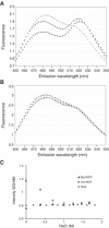
FRET between the two ends of nucleosomal DNA shows that the DNA in Bbd-NCP is less tightly bound. Fluorescence was excited at 385 nm, emission spectra were measured from 450 to 550 nm for mm-NCP (A) and Bbd-NCP (B) at increasing ionic strengths. Only spectra taken at 0, 0.38 and 1 M NaCl are shown (diamonds, squares and triangles, respectively). (C) Ratios of fluorescence intensities (520 nm/480 nm) for mm-NCP, Bbd-NCP and labeled DNA at the indicated salt concentrations (squares, triangles and circles, respectively) are plotted.
Figure 5
Figure 5
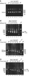
Bbd-NCPs are less resistant against MNase digestion. Identical amounts (2 μg) of mm-146-NCP (A) and Bbd-146-NCP (B) were digested with increasing amounts of MNase (0, 0.05, 0.1, 0.2, 0.4 U) for 1.5 min. Deproteinized samples were analyzed by 10% PAGE, stained with ethidium bromide. For time courses, 10 μg of mm-196-NCP (C) and Bbd-196-NCP (D) was incubated with the same amount of MNase (0.15 U) for the indicated times. In all, 2 μg of sample was removed at 0, 2, 4, 8 and 16 min, and the deproteinized samples were analyzed by 10% PAGE. The 146 bp 5sDNA (146), 196 bp 5sDNA (196) and mixtures of 146 and 73 bp α-satellite DNA (146+73) were loaded as controls, as indicated. DNA size markers are given.
Figure 6
Figure 6
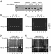
Xla H2A106-NCP and Xla H2A112-NCP behave like wild-type mm-NCP. (A) Salt gradient reconstituted Bbd-NCP (lane 1), Xla-NCP (lanes 6 and 7), Xla H2A106-NCP (106NCP, lanes 2 and 3) and Xla H2A112-NCP (112NCP, lanes 4 and 5), before (−) and after (+) a 1 h incubation at 37°C, were analyzed by 5% native PAGE, followed by staining with Coomassie blue. (B–E) Micrococcal digestion of mutant nucleosomes: 10 μg of Xla-NCP (B), Bbd-NCP (C), Xla H2A112-NCP (D) and Xla H2A106-NCP (E) were digested with 0.15 U MNase. Aliquots of 2 μg were removed after 0, 2, 4, 8 and 16 min. PAGE (10%) was used to check the deproteinized DNA fragments. 5SDNA (146 bp) (146) was loaded as indicated (D and E, lane 8).
Figure 7
Figure 7
The H2A.Bbd docking domain is responsible for the relaxed structure of Bbd-NCP. (A) Salt gradient reconstituted Xla-NCP (lanes 1 and 2), Xla-NCP reconstituted from H2A–H2B dimer and (H3–H4)2 tetramer (X(d+t)-NCP; lane 3), Xla H2A/Bbd-NCP (XB-NCP, lanes 4 and 9), mm-NCP (lanes 5 and 6) and Bbd-NCP (lanes 7 and 8), before (−) and after (+) 1 h incubation at 37°C, were analyzed by 5% native PAGE, stained by Coomassie brilliant blue. (B) Analysis by FRET. Ratios of fluorescence intensity at 520 and 480 nm for Xla-NCP, XB-NCP and labeled DNA at the indicated salt concentrations (squares, triangles and circles, respectively) are plotted. Data points were taken from spectra similar to those shown in Figure 4A and B.
Figure 8
Figure 8
Bbd-containing nucleosomal arrays repress basal transcription. (A) In all, 2.1 μg of assembled Bbd-, mouse- and Drosophila-nucleosomal arrays was digested with 0.12 U MNase. Aliquots of 0.5 μg were removed after 1, 2, 4 and 8 min, deproteinized and analyzed on 1.2% agarose gels. (B) In vitro transcription reactions for naked DNA, Bbd-nucleosomal arrays and mouse-nucleosomal arrays are shown. Tax/CREB and p300 were added as indicated above the lanes. Recovery standard and transcript are indicated on the left. (C) Results from three transcription assays (from three independent chromatin assembly reactions) were quantified by ImageQuant 5.1 and normalized compared to the recovery standard. Transcripts from mouse and Bbd-nucleosomal arrays are shown by white and gray bars, respectively.

References

    1. Akey CW, Luger K (2003) Histone chaperones and nucleosome assembly. Curr Opin Struct Biol 13: 6–14 - PubMed
    1. Anderson MG, Scoggin KE, Simbulan-Rosenthal CM, Steadman JA (2000) Identification of poly(ADP-ribose) polymerase as a transcriptional coactivator of the human T-cell leukemia virus type 1 Tax protein. J Virol 74: 2169–2177 - PMC - PubMed
    1. Becker PB, Horz W (2002) ATP-dependent nucleosome remodeling. Annu Rev Biochem 71: 247–273 - PubMed
    1. Belotserkovskaya R, Oh S, Bondarenko VA, Orphanides G, Studitsky VM, Reinberg D (2003) FACT facilitates transcription-dependent nucleosome alteration. Science 301: 1090–1093 - PubMed
    1. Brooks W, Jackson V (1994) The rapid transfer and selective association of histones H2A and H2B onto negatively coiled DNA at physiological ionic strength. J Biol Chem 269: 18155–18166 - PubMed

Publication types

MeSH terms