. 2004 Aug 4;23(15):3122-32.
doi: 10.1038/sj.emboj.7600314. Epub 2004 Jul 15.

Human geminin promotes pre-RC formation and DNA replication by stabilizing CDT1 in mitosis

Affiliations

Human geminin promotes pre-RC formation and DNA replication by stabilizing CDT1 in mitosis

Andrea Ballabeni et al. EMBO J. .

Abstract

Geminin is an unstable inhibitor of DNA replication that negatively regulates the licensing factor CDT1 and inhibits pre-replicative complex (pre-RC) formation in Xenopus egg extracts. Here we describe a novel function of Geminin. We demonstrate that human Geminin protects CDT1 from proteasome-mediated degradation by inhibiting its ubiquitination. In particular, Geminin ensures basal levels of CDT1 during S phase and its accumulation during mitosis. Consistently, inhibition of Geminin synthesis during M phase leads to impairment of pre-RC formation and DNA replication during the following cell cycle. Moreover, we show that inhibition of CDK1 during mitosis, and not Geminin depletion, is sufficient for premature formation of pre-RCs, indicating that CDK activity is the major mitotic inhibitor of licensing in human cells. Taken together with recent data from our laboratory, our results demonstrate that Geminin is both a negative and positive regulator of pre-RC formation in human cells, playing a positive role in allowing CDT1 accumulation in G2-M, and preventing relicensing of origins in S-G2.


Figures

Figure 1
Figure 1
Human Geminin regulates CDT1 protein levels. (A) Decrease in CDT1 protein levels upon interference with Geminin expression. U2OS cells were either nontransfected or transfected for 60 h with siRNAs to Geminin, CDT1 or unrelated control. Expression of CDT1 and Geminin was determined by immunoblotting together with vinculin as loading control. (B) Two different siRNA target sequences on the gene for human Geminin were tested as in (A) and the expression of the indicated proteins was determined by Western blotting. (C) Expression of a form of Geminin mutated in the target sequence rescues CDT1 protein levels. The decrease in CDT1 levels is specifically due to the loss of Geminin expression. Cells were treated and analyzed as in (A). Control U2OS cells are indicated as ‘−'. HA-Geminin migrates slower than endogenous Geminin.
Figure 2
Figure 2
Human Geminin stabilizes basal levels of CDT1 in S phase. (A) Experimental outline. (B, C) Geminin depletion in S phase leads to a decrease in CDT1 protein levels. (B) U2OS and (C) HeLa were treated during G1 with siRNAs for the indicated genes and collected during S phase 14 and 12 h after nocodazole release, respectively, and treated with DMSO or MG132 for the last 4 h of incubation. The cell lysates were immunoblotted for the indicated proteins. (D) GBD is located between residues 150 and 190 of human CDT1 protein. (E, F) Overexpression of Geminin in S phase stabilizes CDT1 protein. (E) Plasmids expressing HA-CDT1 (WT and ΔGBD) were injected, with or without an expression plasmid for Geminin, in S phase-synchronized HeLa cells and 3 h later fixed and stained for HA or DAPI. IgG were co-injected and counterstained to identify the injected cells. MG132 was used in parallel as control for protein expression. Representative fields are shown. (F) Histogram representing the percentage of cells positive for CDT1 staining in the injected population of cells for each of the treatment shown in (E).
Figure 3
Figure 3
CDT1 accumulation during mitosis is mediated by interaction with Geminin. (A) Outline of the experimental protocol used to transfect G2/M cells with siRNAs (see text for details). (B) Depletion of Geminin in mitosis destabilizes CDT1. U2OS cells were transfected with siRNAs for the indicated genes and immunoblotted for the indicated proteins. ‘As' represents asynchronous cells. The inset shows immunoprecipitated CDT1 incubated with or without alkaline phosphatase (AP) to evaluate the change in electrophoretic mobility. Immunoblotting for Geminin was performed using a 12% acrylamide gel. (C) Indirect immunofluorescence of U2OS cells treated as in (B) and stained for the indicated proteins or DAPI. (D) The CBD is located between residues 112 and 118 of human Geminin, in the first heptad repeat of the coiled-coil (c.c.) domain. (E) Overexpression of a form of myc-Geminin mutated in the target sequence rescues CDT1 protein levels. Depletion of endogenous Geminin in U2OS-overexpressing myc-tagged ΔiGeminin or ΔiGeminin ΔCBD treated as in (B) and immunoblotted for the indicated proteins is shown. (F) MG132 treatment rescues CDT1 protein levels. U2OS cells treated as in (B) to which MG132 was added 4 h before harvesting and immunoblotted for the indicated proteins are shown.
Figure 4
Figure 4
Geminin inhibits CDT1 ubiquitination. (A) myc-Geminin overexpression decreases CDT1 ubiquitination. 293T cells were transfected and synchronized in S phase to detect CDT1 ubiquitination. The upper part of the panel shows the overexpressed proteins for each type of treatment and the use of MG132 for the last 6 h of incubation after release from the thymidine block. In the lower part, immunoblottings for the indicated proteins are shown both for cell lysates and immunoprecipitated samples. Mock immunoprecipitation was performed with an unrelated antibody and is indicated. Myc-Geminin forms (WT and ΔCBD) have lower mobility in comparison to endogenous Geminin. Ubiquitinated forms of CDT1 appear as smeared bands with lower mobility on gel in comparison to endogenous CDT1. (B) myc-Geminin overexpression does not decrease general ubiquitination. Immunoblotting for HA of the same lysates and immunoprecipitated samples indicated in (A) and run on a 17% polyacrylamide gel is shown. The position of the monomeric form of HA-ubiquitin in cell lysates is indicated as well as the smears representing total protein ubiquitination. (C) The inhibition of ubiquitination is not due to secondary cell cycle effects. Immunoblotting for the indicated proteins of the lysates shown in (A) and (B) is represented. ‘As' represents asynchronous and untreated sample.
Figure 5
Figure 5
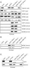
CDK inactivation and not Geminin depletion leads to premature pre-RC formation during mitosis. (A) CDK1 inhibition and not Geminin depletion causes premature MCM loading. U2OS cells treated as in Figure 3B were in parallel treated with roscovitine for the last 3 h of nocodazole incubation and treated in order to separate soluble fraction from chromatin fraction (see Materials and methods for details). The different samples were immunoblotted with antibodies specific for the indicated proteins. ‘sol' indicates the soluble fraction, whereas ‘chrom' indicates the DNaseI-released chromatin fraction. (B) The lack of MCM loading upon Geminin depletion is not due to decrease in CDT1 protein levels. Immunoblotting for the indicated proteins of the same samples shown in (A) or treated with MG132 for the last 3 h of incubation is shown. (C) Geminin binds to both phosphorylated and nonphosphorylated forms of CDT1. Immunoblotting for the indicated proteins (indicated as ‘Input') treated or not with roscovitine, or samples immunoprecipitated with an unrelated antibody or an antibody for Geminin is shown. The differences in mobility on gel are due to roscovitine treatment. MCM2 (in soluble and chromatin fractions) and MCM4 (chromatin fraction) appear as doublets.
Figure 6
Figure 6
Geminin depletion impairs pre-RC formation in the following cell cycle. (A) Outline of the experimental protocol (see text for details). (B) The CDT1 levels in G1 phase depend on the Geminin-mediated mitotic accumulation. The indicated cell lines were treated as in Figure 2B, released from the nocodazole block for 6 h and immunoblotted for the indicated proteins after separation on a 12% polyacrylamide gel. M represents a mitotic sample not released from the nocodazole block. (C) Geminin depletion during mitosis impairs licensing at mitotic exit. U2OS cells treated as in (B) and released into G1 for 6 h were treated in order to separate soluble fraction from chromatin fraction and immunoblotted for the indicated proteins. MCM2 (soluble fraction) appears as a doublet. (D) Geminin and CDT1 depletions during mitosis do not alter mitotic exit rates. FACS analysis profiles of the cells treated as in (B) and (C) and released from the nocodazole block for 1 and 5 h to show the same rate of mitotic exit between the different treatments are shown. (E) Impairment of licensing upon Geminin depletion does not depend on the use of the microtubule-destructing drug nocodazole. Immunoblotting for the indicated proteins of cells treated with the indicated siRNAs during G2 and collected 14 h later without the use of nocodazole (see Materials and methods for details) is shown. (F) Geminin and CDT1 depletions during G2 do not alter cell cycle progression during G2–M–G1 transition. FACS analysis profiles of the samples shown in (E) are represented. The percentages of cells in G1 phase as evaluated by FACS analysis are shown.
Figure 7
Figure 7
Geminin depletion impairs DNA replication in the following cell cycle. (A) Geminin depletion during mitosis impairs DNA replication during the subsequent cell cycle similar to CDT1 depletion. U2OS cells, treated as in Figure 4B, were released from the nocodazole block in the presence of [3H]thymidine and harvested at the indicated time points. The counts per minute (cpm), indicative of [3H]thymidine incorporation, were plotted as mean±s.d. (B) The overexpression of a form of Geminin mutated in the target sequence partially rescues DNA replication. The same protocol as described in (A) was used also for ΔiGeminin-overexpressing cells in parallel with control U2OS (indicated as WT). [3H]thymidine incorporation was evaluated as before 10 h after nocodazole release. The solid bars and striped bars indicate control treatment and siRNA treatment for Geminin, respectively. The insets show immunoblots for chromatin-associated MCM2 and ORC2 in samples prepared 6 h after the nocodazole release.
Figure 8
Figure 8
Model for the role of human Geminin in regulating DNA replication: Geminin promotes licensing at mitotic exit and prevents rereplication during S phase. (A) Human Geminin depletion has different consequences in S and M phases (see Discussion for details). The diagram represents the levels of CDT1 and Geminin during the cell cycle. The two lines are independent lines that do not refer to each other. In the lower part of the panel, U2OS cells synchronized in different ways (see below) were collected at different times during cell cycle and analyzed by FACS analysis or immunoblotted for the indicated proteins. The cells were collected (left to right) 6 h after nocodazole release (lane 1), 4 h (lane 2) and 8 h (lane 3) after thymidine release or taken from the attached (lane 4) or detached (lane 5) populations of nocodazole-treated cells. (B) Human Geminin stabilizes CDT1 during mitosis and appears not to restrict pre-RC formation. In contrast, Geminin is required for the accumulation of CDT1. The main regulator of origin licensing is instead CDK1, which inhibits pre-RC formation by interfering with CDT1 and MCM binding to chromatin.

References

    1. Bastians H, Topper LM, Gorbsky GL, Ruderman JV (1999) Cell cycle-regulated proteolysis of mitotic target proteins. Mol Biol Cell 10: 3927–3941 - PMC - PubMed
    1. Bielinsky AK (2003) Replication origins: why do we need so many? Cell Cycle 2: 307–309 - PubMed
    1. Bousset K, Diffley JF (1998) The Cdc7 protein kinase is required for origin firing during S phase. Genes Dev 12: 480–490 - PMC - PubMed
    1. Cocker JH, Piatti S, Santocanale C, Nasmyth K, Diffley JF (1996) An essential role for the Cdc6 protein in forming the pre-replicative complexes of budding yeast. Nature 379: 180–182 - PubMed
    1. Dahmann C, Diffley JF, Nasmyth KA (1995) S-phase-promoting cyclin-dependent kinases prevent re-replication by inhibiting the transition of replication origins to a pre-replicative state. Curr Biol 5: 1257–1269 - PubMed

Publication types

MeSH terms