

A newly developed peripheral anterior chamber depth analysis system: principle, accuracy, and reproducibility
- PMID: 15258020
- PMCID: PMC1772280
- DOI: 10.1136/bjo.2003.036699
A newly developed peripheral anterior chamber depth analysis system: principle, accuracy, and reproducibility
Abstract
Aim: To develop a new, non-contact system for measuring anterior chamber depth (ACD) quantitatively, and to investigate its accuracy as well as interobserver and intraobserver reproducibility.
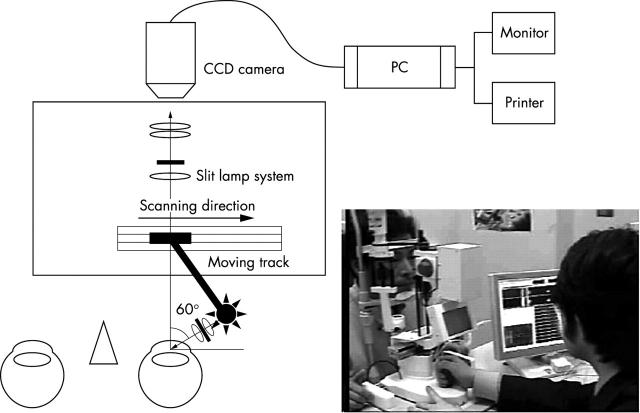
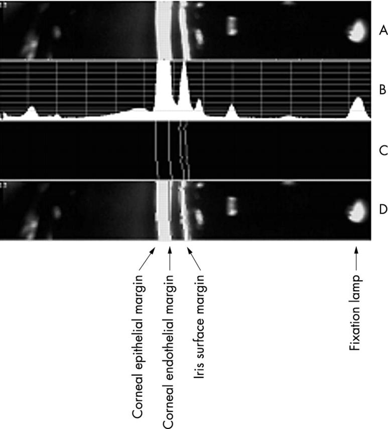
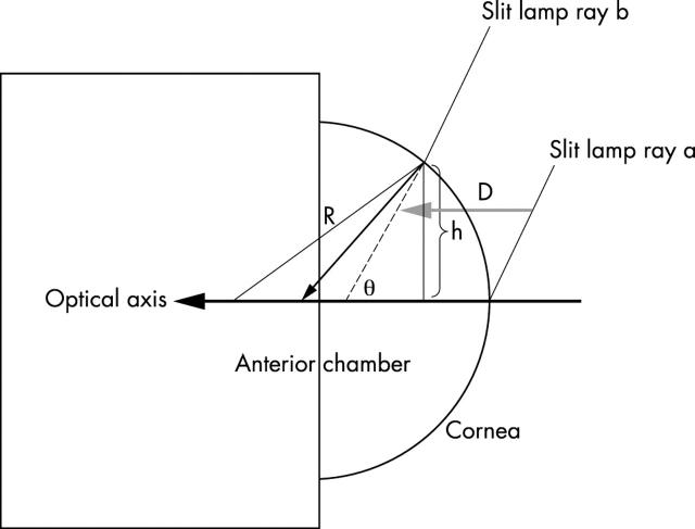
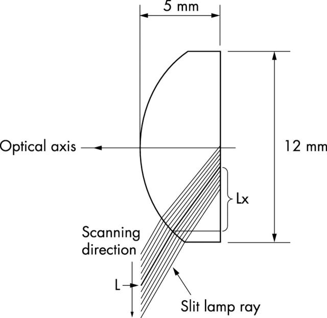
Methods: The system scanned the ACD from the optical axis to the limbus in approximately 0.5 second and took 21 consecutive slit lamp images at 0.4 mm intervals. A computer installed program automatically evaluated the ACD, central corneal thickness (CT), and corneal radius of curvature (CRC) instantly. A dummy eye was used for investigating measurement accuracy. The effects of CT and CRC on the measurement results were examined using a computer simulation model to minimise measurement errors. Three examiners measured the ACD in 10 normal eyes, and interobserver and intraobserver reproducibility was analysed.
Results: The ACD values measured by this system were very similar to theoretical values. Increase of CRC and decrease in CT decreased ACD and vice versa. Data calibration using evaluated CT and CRC successfully reduced measurement errors. Intraobserver and interobserver variations were small. Their coefficient variation values were 7.4% (SD 2.3%) and 6.7% (0.7%), and these values tended to increase along the distance from the optical axis.
Conclusion: The current system can measure ACD with high accuracy as well as high intraobserver and interobserver reproducibility. It has potential use in measuring ACD quantitatively and screening subjects with narrow angle.
Figures


References
-
- Gazzard G , Friedman DS, Devereux JG, et al. A prospective ultrasound biomicroscopy evaluation of changes in anterior segment morphology after laser iridotomy in Asian eyes. Ophthalmology 2003;110:630–8. - PubMed
-
- Van Herick W , Shaffer RN, Schwartz A. Estimation of width of angle of anterior chamber. Incidence and significance of the narrow angle. Am J Ophthalmol 1969;68:62–9. - PubMed
-
- Jacobs IH. Anterior chamber depth measurement using the split-lamp microscope. Am J Ophthalmol 1979;88:236–8. - PubMed
-
- Lee DA, Brubaker RF, Ilstrup DM. Anterior chamber dimensions in patients with narrow angles and angle-closure glaucoma. Arch Ophthalmol 1984;102:46–50. - PubMed
-
- Jin JC, Anderson DR. The effect of iridotomy on iris contour. Am J Ophthalmol 1990;110:260–3. - PubMed
MeSH terms
LinkOut - more resources
Full Text Sources