Review
. 2004 Jul;17(3):495-508, table of contents.
doi: 10.1128/CMR.17.3.495-508.2004.

Emergent human pathogen simian virus 40 and its role in cancer

Affiliations
Review

Emergent human pathogen simian virus 40 and its role in cancer

Regis A Vilchez et al. Clin Microbiol Rev. 2004 Jul.

Abstract

The polyomavirus simian virus 40 (SV40) is a known oncogenic DNA virus which induces primary brain and bone cancers, malignant mesothelioma, and lymphomas in laboratory animals. Persuasive evidence now indicates that SV40 is causing infections in humans today and represents an emerging pathogen. A meta-analysis of molecular, pathological, and clinical data from 1,793 cancer patients indicates that there is a significant excess risk of SV40 associated with human primary brain cancers, primary bone cancers, malignant mesothelioma, and non-Hodgkin's lymphoma. Experimental data strongly suggest that SV40 may be functionally important in the development of some of those human malignancies. Therefore, the major types of tumors induced by SV40 in laboratory animals are the same as those human malignancies found to contain SV40 markers. The Institute of Medicine recently concluded that "the biological evidence is of moderate strength that SV40 exposure could lead to cancer in humans under natural conditions." This review analyzes the accumulating data that indicate that SV40 is a pathogen which has a possible etiologic role in human malignancies. Future research directions are considered.


Figures

FIG. 1.
FIG. 1.
Genetic map of SV40. The circular SV40 DNA genome is represented, with the unique EcoRI site shown at map unit 100/0. Nucleotide numbers based on reference strain SV40-776 begin and end at the origin (Ori) of viral DNA replication (map unit 0/5243). The open reading frames that encode viral proteins are indicated. The early T-ag proteins are shown on the right, and the late structural (VP) proteins are shown on the left. The beginning and end of each open reading frame are indicated by nucleotide numbers. From reference ; used with permission.
FIG. 2.
FIG. 2.
Functional domains of SV40 large T-ag. Known T-ag functions are identified in boxes above and below the shaded bar, which represents the T-ag protein. The numbers given are amino acid residues based on reference strain SV40-776. The variable domain at the extreme C terminus contains amino acid differences among viral strains and is used for strain identification. From reference ; used with permission.
FIG. 3.
FIG. 3.
SV40 large T-ag variable domains. (a) Schematic of large T-ag, showing the location of the variable domain. (b) Amino acid changes in the T-ag C-terminal variable domains of representative SV40 isolates and human primary brain tumor-associated sequences, compared to that of SV40-776. The rectangular boxes represent the T-ag C-terminal region from amino acid (aa) 622 to 708. Virus isolates from monkey kidney cells are shown in the left column. Human brain isolates and primary brain cancer-associated sequences are in the right column. The numbering is according to the system for SV40-776. Del., deletion; Ins., insertion. Arrows indicate the positions and types of amino acid changes. From reference ; used with permission.
FIG. 4.
FIG. 4.
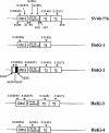
DNA sequence profiles of SV40 regulatory regions detected in human kidney transplant recipients. Ori, viral origin of DNA replication region, which spans nucleotides 5195 to 31; 21-bp repeats, GC-rich region between nucleotides 40 and 103; 72, 72-bp sequence within the enhancer region that is duplicated in some monkey strains (e.g., reference strain SV40-776). Nucleotide numbers are based on SV40-776. Shown are viral sequences associated with transplanted human kidneys (clone designations are on the right). Polymorphisms at the indicated residues are indicated above the boxes. From reference ; used with permission. For a detailed description of the SV40 regulatory region, see reference .
FIG. 5.
FIG. 5.
Regulatory region of SV40. DNA sequence profiles of regulatory regions of SV40 isolates from monkeys and humans and of human tumor-associated DNAs are shown. The diagrams are labeled as described in the legend to Fig. 4. Shown are laboratory-adapted strains (SV40-776, Baylor, and VA45-54), human isolates (SVCPC/SVMEN and SVPML-1), and viral sequences found associated with human brain (CPT, CPP, CPC, and Ep) and bone (Ost) tumors. Tumor-associated sequences usually contain a simple (archetypal) regulatory region without duplications in the enhancer region. From reference ; used with permission.
FIG. 6.
FIG. 6.
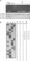
Detection of SV40 T-ag DNA in NHLs. (A) PCR-amplified polyomavirus sequences after agarose gel electrophoresis and staining with ethidium bromide (upper panel) and after Southern blotting with oligonucleotide probes specific for individual polyomaviruses (lower panels). Lane M, molecular weight marker. Positive control reactions included plasmids specific for SV40 (SV40+), JCV (JCV+), and BKV (BKV+). The negative control (NC) was a reaction without added DNA template. CA, cancer control samples (colon and breast cancers). Hybridization of the PCR products to an oligonucleotide probe specific for SV40 identified the lymphoma-associated viral sequences as SV40 specific; JCV and BKV were not detected. (B) Sequence analysis of the PCR product from the N terminus of the T-ag gene. The sequence from the lymphoma sample (NHL) is identical to that of SV40 and distinguishable from those of JCV and BKV. From reference ; used with permission.
FIG. 7.
FIG. 7.
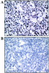
(A) Immunohistochemical detection of SV40 T-ag in a diffuse large B-cell NHL. (B) Negative control specimen (a reactive lymph node). Specimens were stained by using a monoclonal antibody, PAb416. Note the strong nuclear staining of the majority of lymphoma cells. Magnification, ×400.

References

    1. Arrington, A. S., and J. S. Butel. 2001. SV40 and human tumors, p. 461-489. In K. Khalili and G. L. Stoner (ed.), Human polyomaviruses: molecular and clinical perspectives. Wiley-Liss, Inc., New York, N.Y.
    1. Ashkenazi, A., and J. L. Melnick. 1962. Induced latent infection of monkeys with vacuolating SV-40 papova virus: virus in kidneys and urine. Proc. Soc. Exp. Biol. Med. 111:367-372. - PubMed
    1. Atwood, W. J. 2001. Cellular receptors for the polyomaviruses, p. 179-196. In K. Khalili and G. L. Stoner (ed.), Human polyomaviruses: molecular and clinical perspectives. Wiley-Liss, Inc., New York, N.Y.
    1. Atwood, W. J., and L. C. Norkin. 1989. Class I major histocompatibility proteins as cell surface receptors for simian virus 40. J. Virol. 63:4474-4477. - PMC - PubMed
    1. Bergsagel, D. J., M. J. Finegold, J. S. Butel, W. J. Kupsky, and R. L. Garcea. 1992. DNA sequences similar to those of simian virus 40 in ependymomas and choroid plexus tumors of childhood. N. Engl. J. Med. 326:988-993. - PubMed

Publication types

MeSH terms

LinkOut - more resources