. 2004 Jul 27;101(30):11135-40.
doi: 10.1073/pnas.0403925101. Epub 2004 Jul 16.

Genomic DNA double-strand breaks are targets for hepadnaviral DNA integration

Affiliations

Genomic DNA double-strand breaks are targets for hepadnaviral DNA integration

Colin A Bill et al. Proc Natl Acad Sci U S A. .

Erratum in

  • Proc Natl Acad Sci U S A. 2004 Oct 19;101(42):15271

Abstract

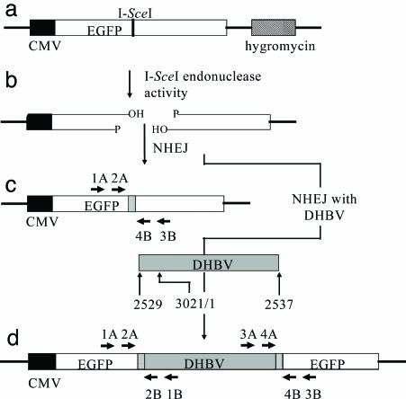
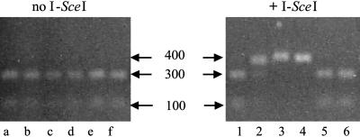
Integrated hepadnaviral DNA in livers and tumors of chronic hepatitis B patients has been reported for many years. In this study, we investigated whether hepatitis B virus DNA integration occurs preferentially at sites of cell DNA damage. A single I-SceI homing endonuclease recognition site was introduced into the DNA of the chicken hepatoma cell line LMH by stable DNA transfection, and double-strand breaks were induced by transient expression of I-SceI after transfection of an I-SceI expression vector. Alteration of the target cleavage site by imprecise nonhomologous end joining occurred at a frequency of approximately 10(-3) per transfected cell. When replication of an avian hepadnavirus, duck hepatitis B virus, occurred at the time of double-strand break repair, we observed integration of viral DNA at the site of the break with a frequency of approximately 10(-4) per transfected cell. Integration depended on the production of viral double-stranded linear DNA and the expression of I-SceI, and integrated DNA was stable through at least 17 cell divisions. Integration appeared to occur through nonhomologous end joining between the viral linear DNA ends and the I-SceI-induced break, because small deletions or insertions were observed at the sites of end joining. The results suggest that integration of hepadnaviral DNA in infected livers occurs at sites of DNA damage and may indicate the presence of more widespread genetic changes beyond that caused by viral DNA integration itself [corrected].


Figures

Fig. 1.
Fig. 1.
Substrate and potential products of NHEJ. (a) Integration target site present in LMH 3.2 cells, consisting of an I-SceI 18-bp recognition sequence inserted into an EGFP gene and the hygromycin-resistance gene for selection. (b) A double-strand break formed by I-SceI endonuclease activity. (c) Product formed by NHEJ of a double-strand break. Precise joining would recreate the I-SceI site, whereas imprecise NHEJ can result in deletions or insertions with concomitant loss of the recognition sequence (gray box). EGFP-specific primer sets 1A/3B and 2A/4B were used to amplify products (see Fig. 2). (d) Product formed by NHEJ, resulting in the integration of DHBV at the double-strand break. The hypothetical DHBV integration substrate shown represents the larger-than-genome size, in situ primed linear DNA, which is the major form of linear DHBV. Integration can be associated with small deletions or insertions of sequence (gray boxes). Left EGFP/DHBV junctions were amplified by nested PCR of genomic DNA by using the primer pairs 1A/1B followed by 2A/2B. Right EGFP/DHBV junctions were amplified similarly from the same genomic DNA by using primers 3A/3B and then 4A/4B.
Fig. 2.
Fig. 2.
Examples of products formed after imprecise joining of double-strand break and repair by NHEJ. An I-SceI expression vector was electroporated into LMH 3.2 cells (+I-SceI) or untransfected (no I-SceI) and incubated for 3 days. Genomic DNA was isolated, digested with I-SceI restriction enzyme to enrich in altered recognition sites, and amplified by PCR using EGFP-specific primer sets 1A/3B and 2A/4B (see Fig. 1 and Table 1). The PCR product was incubated with I-SceI to cleave wild-type products, electrophoresed through a 1.3% agarose gel and stained with ethidium bromide. Two bands indicate that the PCR product still contained the I-SceI recognition sequence (lanes a–f, 1, 5, and 6), and one band indicates loss of this sequence (lanes 3 and 4). Single base changes within the recognition sequence can result in partial digests (lane 2). The sizes of the fragments are given in base pairs.
Fig. 3.
Fig. 3.
Southern blot and genotype analyses of DHBV replicative intermediates. I-SceI expression vector, wild-type 1165A, and 1165A/DR1-13 mutant plasmids were cotransfected into LMH 3.2 cells. After 1, 3, and 6 days of incubation, replicative intermediates were isolated, electrophoresed through a 1.3% agarose gel, transferred to a nylon membrane, and detected by hybridization with a riboprobe specific for detection of the minus strand. The three major forms of replicative intermediates are indicated: rc, relaxed circular DNA; lin, linear double-stranded DNA; ss, single-stranded DNA. The ratios of relaxed circular and linear double-strand DNA (rc/lin) were determined by phosphorimaging. In addition, viral sequences (nucleotides 2669–2840) were amplified by PCR and directly sequenced to determine the ratios of 1165A/1165A-DR1-13 (wt/mut) at five different sites of single-nucleotide polymorphism between the two strains at nucleotide positions 2736, 2742, 2751, 2762, and 2790.
Fig. 4.
Fig. 4.
Agarose gel analysis of left EGFP/DHBV junctions after multiple cell transfers. LMH 3.2 cells were transfected with I-SceI expression vector and 1165A/DR1-13 plasmid and incubated for 3 days (transfer 0) or split ≈1:10 to keep cells in logarithmic growth until 27 days (transfer 5) posttransfection. Genomic DNA was isolated and nested PCR performed as described in the Fig. 1 legend. PCR products were electrophoresed through a 1.3% agarose gel and visualized by ethidium-bromide staining. Molecular marker lanes (m) are included.

References

    1. Beasley, R. P. (1988) Cancer 61, 1942–1956. - PubMed
    1. Levy, L., Renard, C. A., Wei, Y. & Buendia, M. A. (2002) Ann. N.Y. Acad. Sci. 963, 21–36. - PubMed
    1. Birrer, R. B., Birrer, D. & Klavins, J. V. (2003) Ann. Clin. Lab. Sci. 33, 39–54. - PubMed
    1. Summers, J. & Mason, W. S. (1982) Cell 29, 403–415. - PubMed
    1. Brechot, C., Hadchouel, M., Scotto, J., Fonck, M., Potet, F., Vyas, G. N. & Tiollais, P. (1981) Proc. Natl. Acad. Sci. USA 78, 3906–3910. - PMC - PubMed

Publication types

LinkOut - more resources