Expression of cyclooxygenase-2 in human esophageal squamous cell carcinomas
- PMID: 15259059
- PMCID: PMC4724986
- DOI: 10.3748/wjg.v10.i15.2168
Expression of cyclooxygenase-2 in human esophageal squamous cell carcinomas
Abstract
Aim: To determine whether cyclooxygenase-2 (COX-2) was expressed in human esophageal squamous cell carcinoma.
Methods: Quantitative reverse transcription-polymerase chain reaction (RT-PCR), western blotting, immunohistoc-hemistry and immunofluorescence were used to assess the expression level of COX-2 in esophageal tissue.
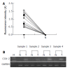
Results: COX-2 mRNA levels were increased by >80-fold in esophageal squamous cell carcinoma when compared to adjacent noncancerous tissue. COX-2 protein was present in 21 of 30 cases of esophageal squamous cell carcinoma tissues, but was undetectable in noncancerous tissue. Immunohistochemistry was performed to directly show expression of COX-2 in tumor tissue.
Conclusion: These results suggest that COX-2 may be an important factor for esophageal cancer and inhibition of COX-2 may be helpful for prevention and possibly treatment of this cancer.
Figures


Similar articles
-
Up-regulation of cyclooxygenase-2 in squamous carcinogenesis of the esophagus.Clin Cancer Res. 2000 Apr;6(4):1229-38. Clin Cancer Res. 2000. PMID: 10778945
-
Dietary zinc modulation of COX-2 expression and lingual and esophageal carcinogenesis in rats.J Natl Cancer Inst. 2005 Jan 5;97(1):40-50. doi: 10.1093/jnci/dji006. J Natl Cancer Inst. 2005. PMID: 15632379
-
Effects of platelet activating factor, butyrate and interleukin-6 on cyclooxygenase-2 expression in human esophageal cancer cells.Scand J Gastroenterol. 2002 Apr;37(4):467-75. doi: 10.1080/003655202317316114. Scand J Gastroenterol. 2002. PMID: 11989839
-
Molecular pathology of cyclooxygenase-2 in neoplasia.Ann Clin Lab Sci. 2000 Jan;30(1):3-21. Ann Clin Lab Sci. 2000. PMID: 10678579 Review.
-
New insights into the functions of Cox-2 in skin and esophageal malignancies.Exp Mol Med. 2020 Apr;52(4):538-547. doi: 10.1038/s12276-020-0412-2. Epub 2020 Apr 1. Exp Mol Med. 2020. PMID: 32235869 Free PMC article. Review.
Cited by
-
Effects of dietary zinc deficiency on esophageal squamous cell proliferation and the mechanisms involved.World J Gastrointest Oncol. 2021 Nov 15;13(11):1755-1765. doi: 10.4251/wjgo.v13.i11.1755. World J Gastrointest Oncol. 2021. PMID: 34853648 Free PMC article.
-
Tpl2 is a key mediator of arsenite-induced signal transduction.Cancer Res. 2009 Oct 15;69(20):8043-9. doi: 10.1158/0008-5472.CAN-09-2316. Epub 2009 Oct 6. Cancer Res. 2009. PMID: 19808956 Free PMC article.
References
-
- Ishizuka T, Tanabe C, Sakamoto H, Aoyagi K, Maekawa M, Matsukura N, Tokunaga A, Tajiri T, Yoshida T, Terada M, et al. Gene amplification profiling of esophageal squamous cell carcinomas by DNA array CGH. Biochem Biophys Res Commun. 2002;296:152–155. - PubMed
-
- Kajiyama Y, Hattori K, Tomita N, Amano T, Iwanuma Y, Narumi K, Udagawa H, Tsurumaru M. Histopathologic effects of neoadjuvant therapies for advanced squamous cell carcinoma of the esophagus: multivariate analysis of predictive factors and p53 overexpression. Dis Esophagus. 2002;15:61–66. - PubMed
-
- Greenberg ER, Baron JA, Freeman DH, Mandel JS, Haile R. Reduced risk of large-bowel adenomas among aspirin users. The Polyp Prevention Study Group. J Natl Cancer Inst. 1993;85:912–916. - PubMed
-
- Giovannucci E, Egan KM, Hunter DJ, Stampfer MJ, Colditz GA, Willett WC, Speizer FE. Aspirin and the risk of colorectal cancer in women. N Engl J Med. 1995;333:609–614. - PubMed
MeSH terms
Substances
LinkOut - more resources
Full Text Sources
Medical
Research Materials

