

Development of a safe neutralization assay for SARS-CoV and characterization of S-glycoprotein
- PMID: 15262502
- PMCID: PMC7127165
- DOI: 10.1016/j.virol.2004.05.017
Development of a safe neutralization assay for SARS-CoV and characterization of S-glycoprotein
Abstract
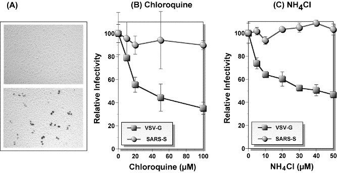
The etiological agent of severe acute respiratory syndrome (SARS) has been identified as a novel coronavirus SARS-CoV. Similar to other coronaviruses, spike (S)-glycoprotein of the virus interacts with a cellular receptor and mediates membrane fusion to allow viral entry into susceptible target cells. Accordingly, S-protein plays an important role in virus infection cycle and is the primary target of neutralizing antibodies. To begin to understand its biochemical and immunological properties, we expressed both full-length and ectodomain of the protein in various primate cells. Our results show that the protein has an electrophoretic mobility of about 160-170 kDa. The protein is glycosylated with high mannose and/or hybrid oligosaccharides, which account for approximately 30 kDa of the apparent protein mass. The detection of S-protein by immunoassays was difficult using human convalescent sera, suggesting that the protein may not elicit strong humoral immune response in virus-infected patients. We were able to pseudotype murine leukemia virus particles with S-protein and produce SARS pseudoviruses. Pseudoviruses infected Vero E6 cells in a pH-independent manner and the infection could be specifically inhibited by convalescent sera. Consistent with low levels of antibodies against S-protein, neutralizing activity was weak with 50% neutralization titers ranging between 1:15 to 1:25. To facilitate quantifying pseudovirus-infected cells, which are stained blue with X-Gal, we devised an automated procedure using an ELISPOT analyzer. The high-throughput capacity of this procedure and the safety of using SARS pseudoviruses should make possible large-scale analyses of neutralizing antibody responses against SARS-CoV.
Figures


Similar articles
-
Evaluation of a novel vesicular stomatitis virus pseudotype-based assay for detection of neutralizing antibody responses to SARS-CoV.J Med Virol. 2006 Dec;78(12):1509-12. doi: 10.1002/jmv.20732. J Med Virol. 2006. PMID: 17063504 Free PMC article.
-
Retroviral vectors pseudotyped with severe acute respiratory syndrome coronavirus S protein.J Virol. 2004 Sep;78(17):9007-15. doi: 10.1128/JVI.78.17.9007-9015.2004. J Virol. 2004. PMID: 15308697 Free PMC article.
-
Receptor-binding domain of severe acute respiratory syndrome coronavirus spike protein contains multiple conformation-dependent epitopes that induce highly potent neutralizing antibodies.J Immunol. 2005 Apr 15;174(8):4908-15. doi: 10.4049/jimmunol.174.8.4908. J Immunol. 2005. PMID: 15814718
-
Expression, glycosylation, and modification of the spike (S) glycoprotein of SARS CoV.Methods Mol Biol. 2007;379:127-35. doi: 10.1007/978-1-59745-393-6_9. Methods Mol Biol. 2007. PMID: 17502675 Free PMC article. Review.
-
Vaccine design for severe acute respiratory syndrome coronavirus.Viral Immunol. 2005;18(2):327-32. doi: 10.1089/vim.2005.18.327. Viral Immunol. 2005. PMID: 16035944 Review.
Cited by
-
High-throughput analysis of anti-poliovirus neutralization antibody titre in human serum by the pseudovirus neutralization test.Sci Rep. 2022 Sep 27;12(1):16074. doi: 10.1038/s41598-022-20544-6. Sci Rep. 2022. PMID: 36167892 Free PMC article.
-
Evaluation of antigenic differences between wild and Sabin vaccine strains of poliovirus using the pseudovirus neutralization test.Sci Rep. 2019 Aug 19;9(1):11970. doi: 10.1038/s41598-019-48534-1. Sci Rep. 2019. PMID: 31427704 Free PMC article.
-
Development of a safe and convenient neutralization assay for rapid screening of influenza HA-specific neutralizing monoclonal antibodies.Biochem Biophys Res Commun. 2010 Jul 2;397(3):580-5. doi: 10.1016/j.bbrc.2010.05.161. Biochem Biophys Res Commun. 2010. PMID: 20617558 Free PMC article.
-
Association between mannose-binding lectin gene polymorphisms and susceptibility to severe acute respiratory syndrome coronavirus infection.J Infect Dis. 2005 Oct 15;192(8):1355-61. doi: 10.1086/491479. Epub 2005 Sep 8. J Infect Dis. 2005. PMID: 16170752 Free PMC article.
-
Development of an enzyme-linked immunosorbent assay-based test with a cocktail of nucleocapsid and spike proteins for detection of severe acute respiratory syndrome-associated coronavirus-specific antibody.Clin Vaccine Immunol. 2009 Feb;16(2):241-5. doi: 10.1128/CVI.00252-08. Epub 2008 Nov 26. Clin Vaccine Immunol. 2009. PMID: 19038782 Free PMC article.
References
-
- Burns J.C., Friedmann T., Driever W., Burrascano M., Yee J.K. Vesicular stomatitis virus G glycoprotein pseudotyped retroviral vectors: concentration to very high titer and efficient gene transfer into mammalian and nonmammalian cells. Proc. Natl. Acad. Sci. U. S .A. 1993;90(17):8033–8037. - PMC - PubMed
-
- Chang S.H., Bae J.L., Kang T.J., Kim J., Chung G.H., Lim C.W., Laude H., Yang M.S., Jang Y.S. Identification of the epitope region capable of inducing neutralizing antibodies against the porcine epidemic diarrhea virus. Mol. Cells. 2002;14(2):295–299. - PubMed
-
- Cho M.W. Subunit protein vaccines: theoretical and practical considerations for HIV-1. Curr. Mol. Med. 2003;3(3):243–263. - PubMed
-
- Cho M.W., Teterina N., Egger D., Bienz K., Ehrenfeld E. Membrane rearrangement and vesicle induction by recombinant poliovirus 2C and 2BC in human cells. Virology. 1994;202(1):129–145. - PubMed
MeSH terms
Substances
Grants and funding
LinkOut - more resources
Full Text Sources
Other Literature Sources
Miscellaneous
