

Review
doi: 10.1503/cmaj.1030055.
Idiopathic pulmonary fibrosis: current understanding of the pathogenesis and the status of treatment
Affiliations
- PMID: 15262886
- PMCID: PMC450366
- DOI: 10.1503/cmaj.1030055
Item in Clipboard
Review
Idiopathic pulmonary fibrosis: current understanding of the pathogenesis and the status of treatment
CMAJ.
.
Abstract
Idiopathic pulmonary fibrosis (IPF) is a progressive and lethal pulmonary fibrotic lung disease. The diagnostic histological changes are called usual interstitial pneumonia and are characterized by histological temporal heterogeneity, whereby normal lung tissue is interspersed with interstitial fibrosis, honeycomb cysts and fibroblast foci. Pulmonary functions show restricted volumes and capacities, preserved flows and evidence of decreased gas exchange. High-resolution computed axial tomography demonstrates evidence of fibrosis and lung remodelling such as honeycomb cysts and traction bronchiectasis. There is no known effective treatment for IPF, but lung transplantation improves survival.
Figures
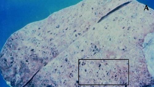
Fig. 1: Gross and histological changes of idiopathic pulmonary fibrosis. A: Section of a lung removed at autopsy from a patient with IPF demonstrates honeycomb cysts. B: Enlargement of the area identified in 1A at a higher magnification shows the cystic lesions of honeycomb cysts. C: Whole-lung thin section highlights the subpleural and basal predominance of pathological changes of IPF, with less involvement of the lung tissue in the more central zones. The pleura is identified by (1); (2) is the preferred site of a diagnostic biopsy, at an interface between honeycomb lung and less involved lung tissue; (3) is an area of honeycomb lung; (4) is a more central area of the lung with minimal changes and appears grossly normal. D: Temporal heterogeneity of histological findings in a single biopsy observed to contain normal lung tissue (N), fibroblast foci (arrows) and interstitial fibrosis (*). Magnification is х40.
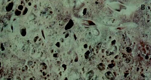
Figure 1. Continued.
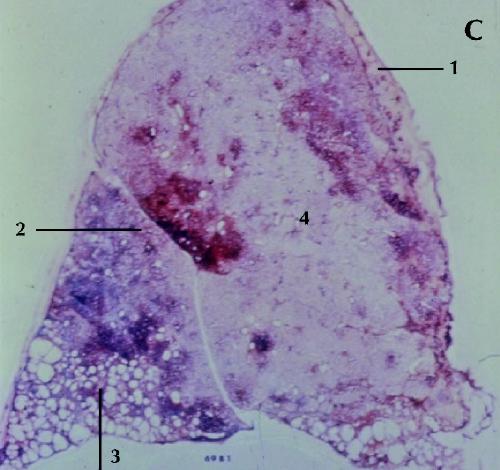
Figure 1. Continued.

Figure 1. Continued.
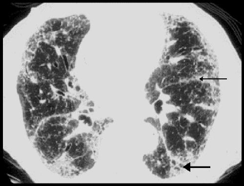
Fig. 2: Radiological changes of IPF on high-resolution CT. Subpleural fibrosis, honeycomb cysts, traction bronchiectasis (thick arrow) and paraseptal fibrosis (thin arrow) are apparent. Relatively minor changes are seen in the central portion of the lung.
References
-
- Katzenstein AL, Myers JL. Nonspecific interstitial pneumonia and the other idiopathic interstitial pneumonias: classification and diagnostic criteria. Am J Surg Pathol 2000;24:1-3. - PubMed
-
- Katzenstein AL, Zisman DA, Litzky LA, Nyuyen BT, Kotloff RM. Usual interstitial pneumonia: histologic study of biopsy and explant specimens. Am J Surg Pathol 2002;26:1567-77. - PubMed
-
- American Thoracic Society. Idiopathic pulmonary fibrosis: diagnosis and treatment. International consensus statement. Am J Respir Crit CareMed 2000; 161: 646-64. - PubMed
-
- American Thoracic Society. ATS/ERS international consensus classification of idiopathic interstitial pneumonias. Am J Respir Crit Care Med 2002; 165: 277-304. - PubMed
-
- Epler GR, Colby TV, McCloud TC, Carrington CB, Gaensler EA. Bronchiolitios obliterans organizing pneumonia. N Engl J Med 1985;312:152-8. - PubMed
Publication types
MeSH terms
LinkOut - more resources
Full Text Sources
Other Literature Sources
Medical