. 2004 Aug;131(15):3581-92.
doi: 10.1242/dev.01227.

Combinatorial Fgf and Bmp signalling patterns the gastrula ectoderm into prospective neural and epidermal domains

Affiliations

Combinatorial Fgf and Bmp signalling patterns the gastrula ectoderm into prospective neural and epidermal domains

Tetsuhiro Kudoh et al. Development. 2004 Aug.

Abstract

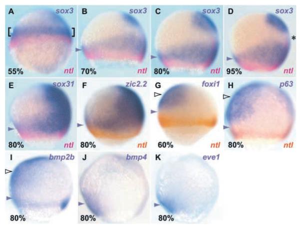
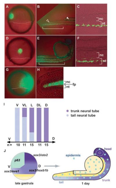
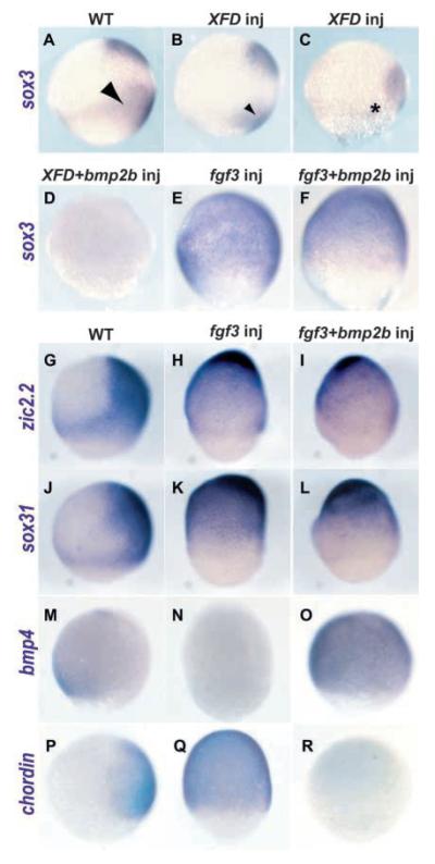
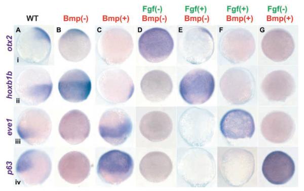
Studies in fish and amphibia have shown that graded Bmp signalling activity regulates dorsal-to-ventral (DV) patterning of the gastrula embryo. In the ectoderm, it is thought that high levels of Bmp activity promote epidermal development ventrally, whereas secreted Bmp antagonists emanating from the organiser induce neural tissue dorsally. However, in zebrafish embryos, the domain of cells destined to contribute to the spinal cord extends all the way to the ventral side of the gastrula, a long way from the organiser. We show that in vegetal (trunk and tail) regions of the zebrafish gastrula, neural specification is initiated at all DV positions of the ectoderm in a manner that is unaffected by levels of Bmp activity and independent of organiser-derived signals. Instead, we find that Fgf activity is required to induce vegetal prospective neural markers and can do so without suppressing Bmp activity. We further show that Bmp signalling does occur within the vegetal prospective neural domain and that Bmp activity promotes the adoption of caudal fate by this tissue.


Figures

Fig. 1
Fig. 1
Vegetal ectoderm expresses prospective neural genes at all DV positions. Lateral views of gastrulae (stage indicated in the bottom left-hand corner) with dorsal towards the right. Genes analysed are indicated on the right of the panels with typeface colour reflecting the colour of the respective expression domains. Vegetal marginal ectoderm is indicated by brackets in A and ventrovegetal ectoderm is indicated by arrowheads in other panels. ntl expression indicates prospective mesoderm (A-H), foxi1 and p63 expression shows prospective epidermis (G,H), which overlaps with the animal domain of bmp2b expression (white arrowheads in G-I). foxi1 expression is initiated earlier in this domain than p63. sox3, sox31 and zic2.2 expression labels all prospective neural tissue (A-F) with the exception, for sox3, of a band of cells in the prospective midbrain and hindbrain (asterisk in D). bmp2b, bmp4 and eve1 are expressed in ventral mesoderm at the blastoderm margin in addition to ventrovegetal ectoderm (purple arrowheads in I-K).
Fig. 2
Fig. 2
Prospective caudal neural tissue is located on the ventral side of the gastrula ectoderm. Lateral views with dorsal towards the right for gastrula stage embryos and to the bottom for 1-day-old embryos. The vegetal limit of the germ ring is indicated by white lines in A, D and G. Gastrula (A,D,G) and 1-day-old (B,C,E,F,H) embryos in which fluorescein was uncaged in ventral vegetal (A) and lateral vegetal (D) ectoderm, or widely throughout dorsal and lateral ectoderm (G), then fates of fluorescent cells were examined the next day. Ventrovegetal cells (A, green fluorescent cells) give rise to tail neural tube (B, bracket; and C in high magnification), whereas laterovegetal ectodermal cells (D, green fluorescent cells) contribute to trunk neural tube (E, bracket; and F in high magnification). Besides neural tissue, some ventrally labelled cells ended up in somite muscle (B, white arrowhead) and in the pronephric duct (B, black arrowhead). In embryos with widespread dorsolateral uncaging of fluorescein (G), labelled cells in the caudal tail are restricted to floor plate, notochord and hypocord, and are absent from more dorsal neural tube (H). (I) Summary of fate-mapping data. Positions at which fluorescein was uncaged in vegetal ectodermal cells are categorised as: V, ventral; VL, ventrolateral, L, lateral; DL, dorsolateral; D, dorsal. The relative contributions of cells to the trunk (rostral to the end of the yolk extension) and tail (caudal to the end of yolk extension) neural tube are indicated by the different colours in each column. The numbers of embryos examined (n) for each position are indicated beneath the columns. (H) Summary of ectodermal expression and fate-mapping data. Ventral ectoderm towards the animal pole generates epidermis/non neural fates, whereas remaining ectoderm generates neural tissue with cells fated to give rise to the most caudal CNS located in ventrovegetal ectoderm and those contributing to the most rostral CNS located in dorsal animal ectoderm (see above) (Kimmel et al., 1990; Woo and Fraser, 1995). The cartoon does not indicate precursors of ventral CNS midline (such as floor plate and hypothalamus) that are positioned close to the organiser (Mathieu et al., 2002; Shih and Fraser, 1996) and distribute widely along the AP axis of the CNS. Indeed, other ventral spinal cord cell types also originate closer to the gastrula organiser than do dorsal spinal cord neurones (Kimmel et al., 1990). Some of the genes selectively expressed in different regions of the ectoderm (see Fig. 7) are indicated. The dorsal (D) and ventral (V) position of cells at gastrula stage does not necessarily correspond with the eventual DV position of the structures to which the cells contribute. For example, spinal cord of the tail (a dorsal structure in 24 hpf embryos) originates from ventrally positioned ectoderm cells of the gastrula [see also recent fate-mapping studies of mesodermal precursors in Xenopus (Kumano and Smith, 2002; Lane and Sheets, 2002)]. fp, floor plate; hp, hypocord; nc, notocord; nt, neural tube.
Fig. 3
Fig. 3
Prospective neural fate in the vegetal ectoderm is initiated independent of the organiser and antagonism of Bmp activity. Lateral views of gastrula stage wild-type (A,F,K), chordin mutant (din−/−) (B,G,L), bmp2b-injected (C,H,M) and ichabod mutant (ich−/−) (D,E,I,J,N,O) embryos with dorsal towards the right (when discernable). Genes analysed by in situ hybridisation are indicated to the left of each row. In E, J and O, embryos were double stained with a probe for ntl, which marks marginal mesoderm cells. The gap (black arrowhead) between ntl and p63 expression domains complementarily expresses sox3 and zic2.2 in ich−/− embryos. The white arrowheads in A-D indicate the position at which sox3 is expressed in prospective forebrain neural tissue in wild-type embryos. Brackets in F-H,K-M indicate the reduction in prospective anterior neural plate size in din−/−, bmp2b-injected and ich−/− embryos.
Fig. 4
Fig. 4
Inhibition of Fgf activity prevents formation of vegetal prospective neural tissue. Lateral views with dorsal right of gastrula stage embryos (80-90% epiboly). Wild-type embryos (A,E,G,I,K) and embryos treated with SU5402, a Fgf receptor inhibitor (B,C,D, with 60 μM SU5402; F,H,J,L, with 90 μM) labelled to show sox3 (A-D), sox31 (E,F) and zix2.2 (G,H) expression, the anterior neural marker gene, otx2 (I,J), and the non neural marker gene, p63 (K,L). The arrowheads in A-D indicate the variable decrease in vegetal sox3 expression and the asterisks show that the animal (anterior) domain of sox3 expression is largely unaffected (although repositioned closer to the vegetal side of the embryo).
Fig. 5
Fig. 5
Fgf can induce prospective neural markers independent of antagonism of Bmp activity. Lateral views with dorsal towards the right (where dorsal is distinguishable) of late gastrula stage wild-type embryos and embryos overexpressing the RNAs indicated above the columns. Genes analysed by in situ hybridisation are indicated to the left of each row. The phenotypes of XFD-expressing embryos are similar to those of SU5402-treated embryos (Fig. 4) with vegetal sox3 expression reduced (A,B, arrowhead; C, asterisk) and animal expression shifted vegetally on the dorsal side of the embryo.
Fig. 6
Fig. 6
Fgf signalling can locally induce sox3 expression and is required for cells to contribute to caudal CNS. (A-L) Views of late gastrula stage wild-type embryos in which donor (d) cells (brown) overexpressing the genes/constructs indicated top right or left side of panels were transplanted at late blastula stage. In C and D, the host (h) embryos are overexpressing the RNAs indicated in the bottom left-hand corner. Genes analysed are indicated in the bottom right-hand corner. Orientation varies depending on the location of the donor cells: (A) laterodorsal, (B) lateral, (C,D) animal, (E,F) dorsal, (G,H) lateral and (I-L) ventral views. In E-L, in situ stained (purple) embryos were photographed before (E,G,I,K) and after (F,H,J,L) staining to reveal donor cells (brown). The domains where sox3 expression was suppressed (E-J) or foxi1 expression was ectopically induced (K,L) are outlined. (M,N) Lateral views of the head (M) and tail (N) of 1-day-old embryos in which XFD-expressing donor cells (d1, green) were mixed and co-transplanted with wild-type donor cells (d2, red) to the same positions in dorsal (M) or ventral (N) vegetal ectoderm at around 50% epiboly stage. In the dorsal transplant, wild-type cells localise to the hindbrain (hb), whereas XFD cells are distributed in the midbrain (mb) and on the surface of the 1-day-old embryo, possibly in the epidermis (epi) (M). In the ventral transplant, wild-type cells localise to tail spinal cord (sp) and somite muscle (mus), whereas XFD-expressing cells are excluded from the spinal cord and localise predominantly to the fin and epidermis (N).
Fig. 7
Fig. 7
Combinatorial activity of Bmp and Fgf signals promotes expression of different regional markers of the gastrula ectoderm. Lateral views of late gastrula stage embryos with dorsal towards the right. Genes analysed are indicated to the left of the rows: otx2, prospective head neural ectoderm; hoxb1b, prospective trunk neural ectoderm; eve1, prospective tail neural ectoderm and mesoderm; p63, prospective epidermis. Bmp activity was modulated by expression of exogenous chordin or bmp2b; Fgf activity was modulated by expression of a dominant-negative Fgf receptor (XFD) or fgf3. The presumed state of activation of the two pathways is indicated in red/green above each column. Note that overexpression of fgf3 (E) induces chordin (see Fig. 2Q) and therefore suppresses Bmp activity (Koshida et al., 2002).
Fig. 8
Fig. 8
Lateral vegetal ectoderm contributes to tail rather than trunk spinal cord in chordino mutant embryos. (A-H) Wild-type (wt), and chordino mutant (din−/−) gastrula stage embryos, with or without injection of fgf3, stained initially to show eve1 expression (red; A,B,E,F) and subsequently to show sox3 expression (purple; C,D,G,H). Arrowheads in A and B indicate the position of vegetal lateral ectoderm, where eve1 expression is absent in wild type (A) while expanded in din−/− (B). The arrowhead in F indicates the dorsal most ectoderm where eve1 expression is absent. (I-N) Lateral views of gastrula (I,L) and 1-day-old (J,K,M,N) wild-type (I-K) and chordin morphant (din MO; L-N) in which fluorescein was uncaged in lateral vegetal ectoderm (I,L, arrowheads) and fates of fluorescent cells were traced on the next day (J,K,M,N). In the wild-type embryo, vegetal lateral ectoderm cells mostly give rise to trunk neural tube (J,K, arrowheads), whereas in the chordin morphant, the cells occupy the tail, including the caudal spinal cord (M,N, arrowheads). White lines in I,L indicate the vegetal limit of the germ ring.
Fig. 9
Fig. 9
Summary of results. The cartoon in the middle represents a lateral view of a gastrula stage embryo. Prospective epidermis is light green; prospective CNS is purple, with dark colour indicating prospective anterior fate and progressively lighter colour indicating progressively more posterior fate (as indicated in the legend to Fig. 2; the organisation of prospective dorsal versus ventral prospective CNS structures is not indicated). Prospective mesendoderm is yellow. Bmp activity is graded during gastrulation: high ventrally and low dorsally (i,ii). This gradient is established, at least in part, by an opposing gradient of dorsally derived Bmp antagonists such as Chordin (e.g. Hammerschmidt and Mullins, 2002). Fgf may also have graded activity during blastula and gastrula stages: high vegetally and low at the animal pole (iii,iv) (Figs 2, 3) (Furthauer et al., 2002; Roehl and Nusslein-Volhard, 2001; Tsang et al., 2002). In the animal gastrula ectoderm, high levels of Bmp activity promotes epidermal fate, while Bmp antagonists promote neural fate (i). Conversely, in vegetal ectoderm, Bmp activity promotes caudal neural fate, whereas Bmp antagonists promote the adoption of more rostral neural fate (ii). The different consequences of activation or suppression of Bmp activity in animal and vegetal ectoderm are influenced by Fgf-dependent promotion of prospective neural fate in vegetal ectoderm (iii). In addition to promoting neural specification, Fgf signalling posteriorises neural tissue in the vegetal ectoderm, most obviously on the dorsal side (iv).

References

    1. Agathon A, Thisse C, Thisse B. The molecular nature of the zebrafish tail organizer. Nature. 2003;424:448–452. - PubMed
    1. Akai J, Storey K. Brain or brawn: how FGF signaling gives us both. Cell. 2003;115:510–512. - PubMed
    1. Amacher SL, Draper BW, Summers BR, Kimmel CB. The zebrafish T-box genes no tail and spadetail are required for development of trunk and tail mesoderm and medial floor plate. Development. 2002;129:3311–3323. - PubMed
    1. Amaya E, Musci TJ, Kirschner MW. Expression of a dominant negative mutant of the FGF receptor disrupts mesoderm formation in Xenopus embryos. Cell. 1991;66:257–270. - PubMed
    1. Aybar MJ, Mayor R. Early induction of neural crest cells: lessons learned from frog, fish and chick. Curr. Opin. Genet. Dev. 2002;12:452–458. - PubMed

Publication types

MeSH terms