. 2004 Aug;186(15):5062-77.
doi: 10.1128/JB.186.15.5062-5077.2004.

The homogentisate pathway: a central catabolic pathway involved in the degradation of L-phenylalanine, L-tyrosine, and 3-hydroxyphenylacetate in Pseudomonas putida

Affiliations

The homogentisate pathway: a central catabolic pathway involved in the degradation of L-phenylalanine, L-tyrosine, and 3-hydroxyphenylacetate in Pseudomonas putida

Elsa Arias-Barrau et al. J Bacteriol. 2004 Aug.

Abstract

Pseudomonas putida metabolizes Phe and Tyr through a peripheral pathway involving hydroxylation of Phe to Tyr (PhhAB), conversion of Tyr into 4-hydroxyphenylpyruvate (TyrB), and formation of homogentisate (Hpd) as the central intermediate. Homogentisate is then catabolized by a central catabolic pathway that involves three enzymes, homogentisate dioxygenase (HmgA), fumarylacetoacetate hydrolase (HmgB), and maleylacetoacetate isomerase (HmgC), finally yielding fumarate and acetoacetate. Whereas the phh, tyr, and hpd genes are not linked in the P. putida genome, the hmgABC genes appear to form a single transcriptional unit. Gel retardation assays and lacZ translational fusion experiments have shown that hmgR encodes a specific repressor that controls the inducible expression of the divergently transcribed hmgABC catabolic genes, and homogentisate is the inducer molecule. Footprinting analysis revealed that HmgR protects a region in the Phmg promoter that spans a 17-bp palindromic motif and an external direct repetition from position -16 to position 29 with respect to the transcription start site. The HmgR protein is thus the first IclR-type regulator that acts as a repressor of an aromatic catabolic pathway. We engineered a broad-host-range mobilizable catabolic cassette harboring the hmgABC, hpd, and tyrB genes that allows heterologous bacteria to use Tyr as a unique carbon and energy source. Remarkably, we show here that the catabolism of 3-hydroxyphenylacetate in P. putida U funnels also into the homogentisate central pathway, revealing that the hmg cluster is a key catabolic trait for biodegradation of a small number of aromatic compounds.


Figures

FIG. 1.
FIG. 1.
Pathway for the catabolism of Phe and Tyr. (A) Arrangement of the genes involved in catabolism of Phe and Tyr in P. putida U. The gene clusters encoding the peripheral and central (homogentisate) pathways are indicated. Discontinuous lines between genes indicate unknown distances. The relative positions of the gene clusters in the genome of P. putida U are still unknown. (B) Biochemistry of the Phe/Tyr catabolism. The intermediates of the catabolic pathway are indicated. The homogentisate central pathway is enclosed in a box. The enzymes are PhhA (phenylalanine hydroxylase), PhhB (carbinolamine dehydratase), TyrB (tyrosine aminotransferase), Hpd (4-OH-PhPy dioxygenase), HmgA (homogentisate dioxygenase), HmgB (fumarylacetoacetate hydrolase), HmgC (maleylacetoacetate isomerase), Mha (3-hydroxyphenylacetate monooxygenase), and dihydropteridine reductase (DHPR). In previous work, TyrB, HmgB, and HmgC were called PhhC, Fah and Mai, respectively.
FIG. 2.
FIG. 2.
Pigment production (browning) by wild-type and mutant P. putida U strains: growth of wild-type P. putida U (a) and the P. putida U-95 mutant strain (b) on MM containing 5 mM Tyr and 4-OH-PhAc (plate 1) or 5 mM 3-OH-PhAc and 4-OH-PhAc (plate 2).
FIG. 3.
FIG. 3.
Structure of 2,5-OH-PhAc.
FIG. 4.
FIG. 4.
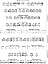
Gene organization of the clusters encoding the homogentisate central pathway and the Phe/Tyr peripheral pathway in P. putida KT2440, and comparisons with equivalent gene clusters from other bacteria. Genes are represented by arrows as follows: black, regulatory genes; stippled, transport genes; vertically striped, genes encoding the homogentisate dioxygenase; hatched, genes encoding the hydrolase and isomerase of the homogentisate pathway; cross-hatched, genes encoding the 4-OH-PhPyr dioxygenase; white, genes encoding the tyrosine aminotransferase; horizontally striped, genes encoding the phenylalanine hydroxylase and carbinolamine dehydratase. The numbers beneath the arrows indicate the levels of amino acid sequence identity (expressed as percentages) between the encoded gene products and the equivalent products from P. putida. Identity values are not shown for the hmgR gene products that do not belong to the IclR family of transcriptional regulators. The genomes of P. fluorescens, A. vinelandii, and S. pomeroyi are not completely assembled.
FIG. 5.
FIG. 5.
Tyr and 3-OH-PhAc share the same central catabolic pathway: growth of wild-type P. putida U (•) and the mutants P. putida U-dhmgA (▪), U-dhmgB (▴), U-dhmgC (▾), U-Δhmg (○), U-Δhmg(pMChmg) (▵) in MM containing 5 mM Tyr (A) or 10 mM 3-OH-PhAc (B) as the sole carbon and energy source.
FIG. 6.
FIG. 6.
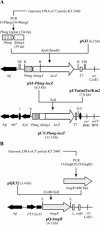
Catabolic cassette for the catabolism of Tyr. (A) Schematic representation of the construction and expression of a Tyr catabolic cassette. The primer pairs used for PCR amplification are shown in Table 2. Genes are indicated by arrows. The Plac and Phmg promoters are shown. Apr and Kmr, genes that confer ampicillin and kanamycin resistance, respectively. The pBBR1 and pUC origins of replication (oripBBR1 and oripUC) are indicated. oriTRP4, RP4-mediated mobilization (Mob+) functions. (B) Growth of recombinant strain E. coli(pU-HHP) (•) and control strain E. coli(pUC18) (▴) in MM supplemented with 0.05% Casamino Acids and 5 mM Tyr as the sole carbon and energy source.
FIG. 7.
FIG. 7.
Scheme for subcloning of the hmg regulatory elements: schematic representation of construction of a Phmg::lacZ translational fusion cassette (A) and of plasmid pQ-hmgR harboring the regulatory hmgR gene (B). DNA fragments were PCR amplified by using primers described in Table 2. The Phmg fragment was cloned into the promoter-probe pSJ3 plasmid. The hmgR fragment was cloned into the pQE32 gene expression vector. ΔhmgA indicates a truncated hmgA gene (the number of amino acid [aa] residues fused to the LacZ protein is shown in parentheses). T7, to, and rrnB, transcriptional terminators from the T7 and lambda phages and T1 transcriptional terminator from the E. coli rrnB operon, respectively; I and O, termini of the mini-Tn5 transposons; Apr and Kmr, genes that confer ampicillin and kanamycin resistance, respectively; tnp*, gene devoid of NotI sites encoding the Tn5 transposase; PT5/lacO, hybrid promoter-operator region composed of the PT5 promoter of phage T5 and the lacO operator from the E. coli lac cluster; oriTRP4, RP4-mediated mobilization functions. The R6K and ColE1 origin of replication (oriR6K and oriColE1) are indicated. Restriction sites: B, BamHI; E, EcoRI; H, HindIII; K, KpnI; N, NotI; S, SalI.
FIG. 8.
FIG. 8.
Analysis of the hmgR-hmgA intergenic region. (A) Identification of the transcription start site in Phmg. Primer extension experiments were carried out by using total RNA isolated from E. coli AF141 cells bearing the lacZ translational fusion plasmid pSJ-Phmg-lacZ (lane 2) and the control plasmid pSJ3 (lane 1). The size of the extended product was determined by comparison with the DNA sequencing ladder of the Phmg promoter region (lanes T, C, G, and A). Primer extension and sequencing reactions were performed with primer O-Phmg3 as described in Materials and Methods. The nucleotide sequence surrounding the transcription initiation site (enclosed in a box) in the coding strand is shown. (B) Schematic representation of regulation of the hmg cluster and nucleotide sequence of the hmgR-hmgA intergenic region. The hmgR regulatory gene is indicated by a thick grey arrow. The hmgABC catabolic genes are indicated by a thick open arrow. The minus sign indicates transcriptional repression by the HmgR protein. The plus sign indicates transcriptional activation (induction) promoted by homogentisate. Homogentisate is transformed into fumarate and acetoacetate by the HmgABC proteins. The nucleotide sequence of the Phmg probe (335 bp) is indicated. The translation initiation codon for the hmgA and hmgR genes is indicated by boldface lowercase letters; the bent arrows indicate the direction of transcription. The transcription start site (position +1) and the inferred −10 and −35 boxes of the Phmg promoter are indicated. The HmgR binding region is indicated by brackets. The repeated motifs are indicated by thin grey arrows. RBS, ribosome binding site.
FIG. 9.
FIG. 9.
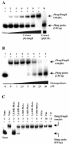
Gel retardation analyses of HmgR binding to the hmgR-hmgA intergenic region. Cell extracts were prepared and gel retardation analyses were performed as described in Materials and Methods. The probe DNA used, Phmg, was PCR amplified from plasmid pSJ-Phmg-lacZ as described in Materials and Methods. (A) Lanes 1 to 7, retardation assay mixtures containing 0, 0.5, 0.7, 1.0, 1.5, 2.0, and 3.0 μg of total protein, respectively, of HmgR+ extracts obtained from cells bearing plasmid pQ-hmgR; lane 8, assay mixture containing 3.0 μg of total protein of HmgR extracts obtained from cells bearing the control plasmid pQE32. (B) Lanes 2 to 8, retardation assay mixtures containing 3.0 μg of total protein of HmgR+ extracts in the absence of 2,5-OH-PhAc (lane 2) or in the presence of increasing concentrations of 2,5-OH-PhAc, as follows: 1 μM (lane 3), 2.5 μM (lane 4), 5.0 μM (lane 5), 10.0 μM (lane 6), 25.0 μM (lane 7), and 50.0 μM (lane 8). Lane 1 shows migration of the Phmg probe without protein extract. (C) Gel retardation assays with 3.0 μg of total protein of HmgR+ extracts and the following different ligands at a concentration of 1 mM: 2,5-OH-PhAc (lane 3), 2,5-OH-benzoate (2,5-OH-Benz) (lane 4), 2-OH-PhAc (lane 5), 3-OH-PhAc (lane 6), 3,4-OH-PhAc (lane 7), 4-OH-PhPyr (lane 8), PhAc (lane 9), Phe (lane 10), and Tyr (lane 11). Lanes 1 and 2 contained assay mixtures lacking HmgR+ extract and ligand, respectively. The positions of DNA probes and the DNA-HmgR complexes are indicated by arrows.
FIG. 10.
FIG. 10.
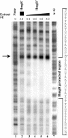
DNase I footprinting analyses of the interaction of HmgR with the Phmg promoter region. The DNase I footprinting experiments were carried out by using the Phmg probe labeled at the 5′ end of the noncoding strand as described in Materials and Methods. Lanes 1 and 3 to 6 contained footprinting assay mixtures containing 0, 0.1, 0.3, 1.0, and 3.0 μg of total protein of HmgR+ extracts (pQ-hmgR), respectively. Lane 2 contained a footprinting assay mixture with 3.0 μg of total protein of HmgR extracts (pQE32). Lane 7 shows the results for the A+G Maxam-Gilbert sequencing reaction (40) that provided the sequence of the Phmg probe. The HmgR protected region is indicated, and an expanded view of the nucleotide sequence is indicated by brackets. The asterisk indicates the transcription initiation site in the Phmg promoter. A DNase I-hypersensitive site is indicated by an arrow.
FIG. 11.
FIG. 11.
Comparison of the Phmg promoter regions in several Pseudomonas species. The hmgR-hmgA intergenic regions from P. putida, P. fluorescens, P. aeruginosa, and P. syringae were aligned from position −50 to position 40 with respect to the transcription initiation site in P. putida. The asterisks indicate the conserved nucleotides. The −35 and −10 boxes, the +1 transcription initiation site, the ribosome binding site (RBS), and the ATG translation initiation codon from hmgA in P. putida are indicated by a black background. The HmgR binding region is indicated by brackets. Repeated motifs are indicated by arrows.

References

    1. Adachi, K., Y. Iwayama, H. Tanioka, and Y. Takeda. 1966. Purification and properties of homogentisate oxygenase from Pseudomonas fluorescens. Biochim. Biophys. Acta 118:88-97. - PubMed
    1. Bayley, S. A., C. J. Duggleby, M. J. Worsey, P. A. Williams, K. G. Hardy, and P. Broda. 1977. Two modes of loss of the Tol function from Pseudomonas putida mt-2. Mol. Gen. Genet. 154:203-204. - PubMed
    1. Boyer, H. W., and D. Roulland-Dussoix. 1969. A complementary analysis of the restriction and modification of DNA in Escherichia coli. J. Mol. Biol. 41:459-472. - PubMed
    1. Bradford, M. M. 1976. A rapid and sensitive method for the quantitation of microgram quantities of protein utilizing the principle of protein-dye binding. Anal. Biochem. 72:248-254. - PubMed
    1. Buell, C. R., V. Joardar, M. Lindeberg, J. Selengut, I. T. Paulsen, M. L. Gwinn, R. J. Dodson, R. T. Deboy, A. S. Durkin, J. F. Kolonay, R. Madupu, S. Daugherty, L. Brinkac, M. J. Beanan, D. H. Haft, W. C. Nelson, T. Davidsen, N. Zafar, L. Zhou, J. Liu, Q. Yuan, H. Khouri, N. Fedorova, B. Tran, D. Russell, K. Berry, T. Utterback, S. E. Van Aken, T. V. Feldblyum, M. D'Ascenzo, W. L. Deng, A. R. Ramos, J. R. Alfano, S. Cartinhour, A. K. Chatterjee, T. P. Delaney, S. G. Lazarowitz, G. B. Martín, D. J. Schneider, X. Tang, C. L. Bender, O. White, C. M. Fraser, and A. Collmer. 2003. The complete genome sequence of the Arabidopsis and tomato pathogen Pseudomonas syringae pv. tomato DC3000. Proc. Natl. Acad. Sci. USA 100:10181-10186. - PMC - PubMed

Publication types

MeSH terms

Associated data

LinkOut - more resources