. 2004 Jul 19;166(2):225-35.
doi: 10.1083/jcb.200402073.

Cortactin and Crk cooperate to trigger actin polymerization during Shigella invasion of epithelial cells

Affiliations

Cortactin and Crk cooperate to trigger actin polymerization during Shigella invasion of epithelial cells

Laurence Bougnères et al. J Cell Biol. .

Abstract

Shigella, the causative agent of bacillary dysentery, invades epithelial cells in a process involving Src tyrosine kinase signaling. Cortactin, a ubiquitous actin-binding protein present in structures of dynamic actin assembly, is the major protein tyrosine phosphorylated during Shigella invasion. Here, we report that RNA interference silencing of cortactin expression, as does Src inhibition in cells expressing kinase-inactive Src, interferes with actin polymerization required for the formation of cellular extensions engulfing the bacteria. Shigella invasion induced the recruitment of cortactin at plasma membranes in a tyrosine phosphorylation-dependent manner. Overexpression of wild-type forms of cortactin or the adaptor protein Crk favored Shigella uptake, and Arp2/3 binding-deficient cortactin derivatives or an Src homology 2 domain Crk mutant interfered with bacterial-induced actin foci formation. Crk was shown to directly interact with tyrosine-phosphorylated cortactin and to condition cortactin-dependent actin polymerization required for Shigella uptake. These results point at a major role for a Crk-cortactin complex in actin polymerization downstream of tyrosine kinase signaling.


Figures

Figure 1.
Figure 1.
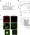
Cortactin dsRNA interference inhibits Shigella entry. HeLa cells were transfected with transfection reagent alone (ctr), sense and antisense strands of dsRNA1, scrambled version of dsRNA1 (scrambled), or dsRNA1. (a) 24 h after transfection, cell lysates were analyzed by Western blot using anti-cortactin (cortactin), anti-Arp3 (arp3), or anti-actin (actin) antibodies. A decrease in the cortactin levels was observed in dsRNA1-transfected cells in three independent experiments. (b–d) 24 h after transfection, cells were challenged with Shigella, fixed, and processed for staining. (b) Bacterial uptake was determined by differential outside/total bacteria immunofluorescence staining in the different controls and in dsRNA1-treated cells. (c) Representative fields of scrambled or dsRNA1-transfected cells challenged with Shigella; bacteria (blue), cortactin (red), and F-actin (green). Bottom panels correspond to higher magnification of the inset in the middle panels. Bars, 10 μm. (d) The frequency of F-actin foci formation was determined for each sample by counting foci in 600 cells. The plotted data were averaged from the scoring of samples from three independent experiments ± SEM.
Figure 2.
Figure 2.
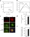
Overexpression of cortactin enhances Shigella-induced actin polymerization and bacterial uptake. HeLa cells were transfected with vector alone (mock) or Flag-tagged cortactin (FL). After 24 h, cells were infected with Shigella, fixed, and processed for fluorescent labeling. Transfected cells were visualized by staining of FL with anti-Flag antibody. (a) Shigella uptake is shown for mock cells (dotted line) and FL cells (solid line). (b) The frequency of Shigella-induced actin foci formation is shown for mock cells (dotted line) and FL cells (solid line). (c) Representative confocal images of Shigella foci observed in mock or FL cells, stained for Flag-tagged cortactin (red), F-actin (green), and Shigella (blue). Bar, 5 μm. (d and e) Quantitative analysis of the surface area and the fluorescence intensity per surface unit of F-actin foci was performed in mock cells (white bar) and FL cells (solid bar), using a dedicated computer program. All the plotted data shown in this figure were averaged from three independent experiments ± SEM.
Figure 3.
Figure 3.
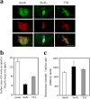
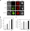
Expression of kinase-inactive Src and TM cortactin inhibits Shigella-induced actin foci formation. (a) Representative confocal microscopy images of Shigella entry structures observed in mock cells (mock), SrcK− cells (SrcK−), and cells expressing tyrosine-mutated cortactin (TM). Cells were challenged for 15 min with Shigella, fixed, and processed for fluorescence labeling of F-actin (green), cortactin (red), and bacterial LPS (blue). Bar, 5 μm. (b and c) Surface area and fluorescence intensity per surface unit of F-actin in Shigella entry structures were quantified in mock, SrcK−, and TM cells from images obtained with an epifluorescence microscope using a dedicated computer program (see Materials and methods). The analysis was performed on 60 foci from three independent experiments.
Figure 4.
Figure 4.
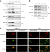
Shigella invasion induces cortactin association with plasma membranes. After challenge with noninvasive (NON INV) or invasive (INV) Shigella, cell extracts were fractionated using a discontinuous sucrose gradient. (a) Extracts from HeLa cells were fractionated and analyzed by anti-cortactin, anti-actin, anti-tubulin, and anti-Crk Western blot. Invasive Shigella induces a shift of cortactin to the plasma membrane–containing fraction. Actin and Crk co-shift with cortactin, but not with tubulin. (b) Extracts from cells transfected with full-length (FL) or tyrosine-mutated cortactin (TM) were fractionated and analyzed by anti-Flag Western blot. (c) Cells were surface labeled with a rhodamine derivative (red) previous to bacterial invasion. After cell extract fractionation on sucrose gradients, the plasma membrane–enriched 46% sucrose fraction was incubated with FITC-fluorescent beads (green) coupled to anti-cortactin or anti-β1 integrin antibodies. Beads were recovered by centrifugation and fluorescence analysis was performed to determine the presence of plasma membranes. Bar, 5 μm.
Figure 5.
Figure 5.
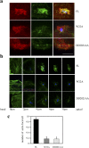
Cortactin-dependent actin polymerization during Shigella entry requires the cortactin–Arp2/3 binding site. (a and b) HeLa cells were transfected with wild-type cortactin (FL), the W22A cortactin mutant, or DD2021AA. 24 h after transfection, cells were challenged 15 min with Shigella, fixed, processed for fluorescence labeling, and analyzed by confocal microscopy. Samples were stained for exogenous cortactin (red), F-actin (green), and anti-LPS (blue). Bar, 5 μm. (b) Confocal planes of Shigella actin foci staining from the basal to apical region spaced by 2 μm. (c) The number of Shigella-induced actin foci per cell was determined from three independent experiments ± SEM. Mutations in the cortactin–Arp2/3 binding site impair the formation of Shigella-induced actin extensions.
Figure 6.
Figure 6.
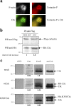
Cortactin associates with Crk during Shigella entry. (a) Cells were challenged 15 min with Shigella, fixed, and processed for fluorescence labeling of F-actin (gray), Crk (green), and tyrosine-phosphorylated cortactin (red). Images were acquired with an epifluorescence microscope. Bar, 5 μm. (b) FL cortactin was immunoprecipitated from HeLa cells coexpressing Flag-tagged cortactin and HA-tagged Crk, previously infected 15 min with noninvasive or invasive Shigella. Immune complexes were separated by SDS-PAGE, transferred to a membrane filter, and the same membrane was immunoblotted using anti-Flag or anti-HA antibodies, followed by ECL+ detection (Amersham Biosciences). For each sample, the relative fold increase corresponds to the intensity of the signal obtained for HA-Crk over that of Flag-cortactin, assigning the value “1” for the sample challenged with the noninvasive strain. (c) Immunoprecipitation from mock-, wtCrk-, or R38VCrk-transfected cell lysates using anti-Crk antibody was followed by gel electrophoresis. Anti-Crk Western blot analysis shows endogenous and recombinant Crk. Overlay analysis was performed using GST submitted to a kinase assay (GST*), GST-cortactin (Cort), or phosphorylated GST-cortactin (Cort-P) as probes. The bold number indicates the normalized value obtained for binding of cortactin or phosphorylated cortactin to bands labeled with an asterisk (see Materials and methods). Phosphorylated cortactin binds to endogenous or recombinant HA-Crk, but not to R38VCrk.
Figure 7.
Figure 7.
Cortactin and Crk cooperate to stimulate the formation of Shigella-induced F-actin–rich extensions. HeLa cells were transfected with vector alone (mock), Myc-tagged wild-type Crk (wtCrk), or FL cortactin (FL), or were cotransfected with Myc-tagged wild-type Crk and Flag-tagged FL cortactin (wtCrk-FL). (a) Representative confocal images of Shigella entry structures observed in mock transfectants, wtCrk transfectants, and wtCrk-FL cortactin cotransfectants. Cells were challenged 15 min with Shigella, fixed, and processed for fluorescence labeling of F-actin (black and white), Myc-Crk (green), and endogenous cortactin (red) in mock and wtCrk cells or of recombinant cortactin (red) in wtCrk-FL transfectants. Differential interference contrast (DIC) images show cell extension morphology at Shigella entry site. Bar, 5 μm. (b and c) Quantitative analysis of the surface area and the fluorescence intensity per surface unit of F-actin foci was performed in mock, wtCrk, FL, or wtCrk-FL transfectants using a dedicated computer program. Plotted data were averaged from three independent experiments ± SEM.
Figure 8.
Figure 8.
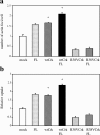
Coupled action of Crk and cortactin is required for efficient uptake of Shigella. HeLa cells were transfected with vector alone (mock), FL cortactin (FL), Myc-tagged wild-type Crk (wtCrk), or HA-tagged mutant Crk (R38VCrk), or were cotransfected with Myc-tagged wild-type Crk and Flag-tagged FL cortactin (wtCrk-FL), HA-tagged mutant Crk, and Flag-tagged FL cortactin (R38VCrk-FL). After 24 h of transfection, cells were infected with Shigella, fixed, and processed for fluorescent labeling. (a) The frequency of Shigella-induced actin foci formation at 15 min was scored for each transfectant. (b) Shigella uptake was determined for each transfectant using outside/total staining. The results of each experiment were normalized to the value obtained for mock transfectants, corresponding to 37% uptake. The average number of cell-associated bacteria was 4.5 bacteria per cell, and did not vary significantly in the different samples. The plotted data were averaged from the scoring of samples from three independent experiments ± SEM. Statistical significance was assessed by t test (denoted by asterisks).

Similar articles

Cited by

References

    1. Abassi, Y.A., and K. Vuori. 2002. Tyrosine 221 in Crk regulates adhesion-dependent membrane localization of Crk and Rac and activation of Rac signaling. EMBO J. 21:4571–4582. - PMC - PubMed
    1. Allaoui, A., P.J. Sansonetti, and C. Parsot. 1992. MxiJ, a lipoprotein involved in secretion of Shigella Ipa invasins, is homologous to YscJ, a secretion factor of the Yersinia Yop proteins. J. Bacteriol. 174:7661–7669. - PMC - PubMed
    1. Bowden, E.T., M. Barth, D. Thomas, R.I. Glazer, and S.C. Mueller. 1999. An invasion-related complex of cortactin, paxillin and PKCμ associates with invadopodia at sites of extracellular matrix degradation. Oncogene. 18:4440–4449. - PubMed
    1. Burton, E.A., R. Plattner, and A.M. Pendergast. 2003. Abl tyrosine kinases are required for infection by Shigella flexneri. EMBO J. 22:5471–5479. - PMC - PubMed
    1. Cheresh, D.A., J. Leng, and R.L. Klemke. 1999. Regulation of cell contraction and membrane ruffling by distinct signals in migratory cells. J. Cell Biol. 146:1107–1116. - PMC - PubMed

Publication types

MeSH terms