. 2004 Jul 19;166(2):249-60.
doi: 10.1083/jcb.200312054.

Synaptotagmins are trafficked to distinct subcellular domains including the postsynaptic compartment

Affiliations

Synaptotagmins are trafficked to distinct subcellular domains including the postsynaptic compartment

Bill Adolfsen et al. J Cell Biol. .

Abstract

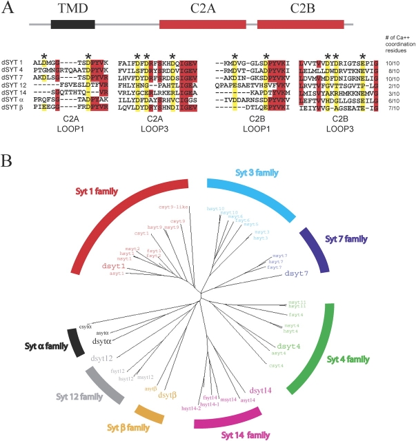
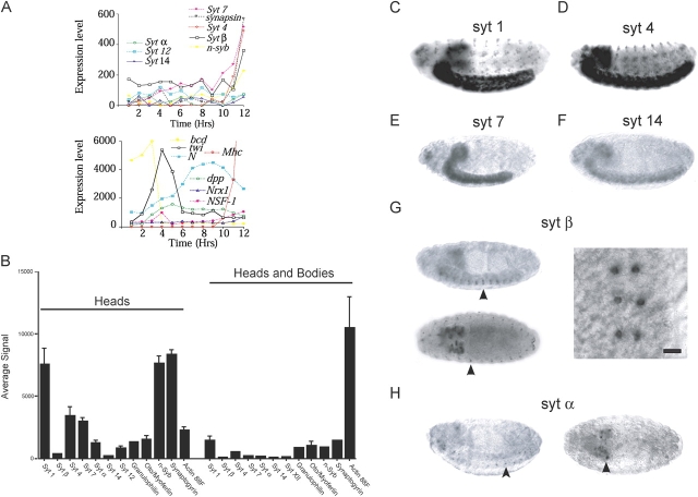
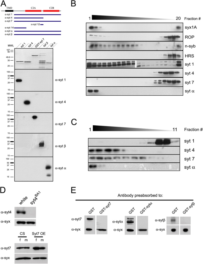
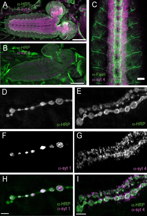
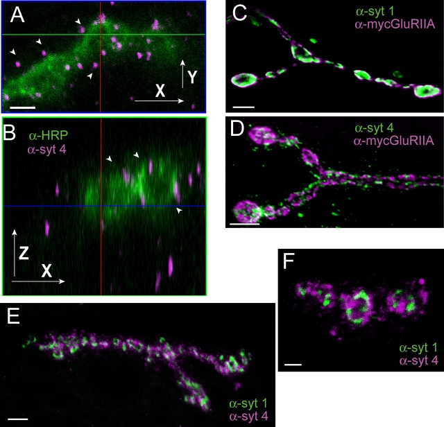
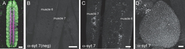
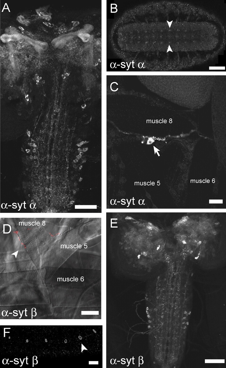
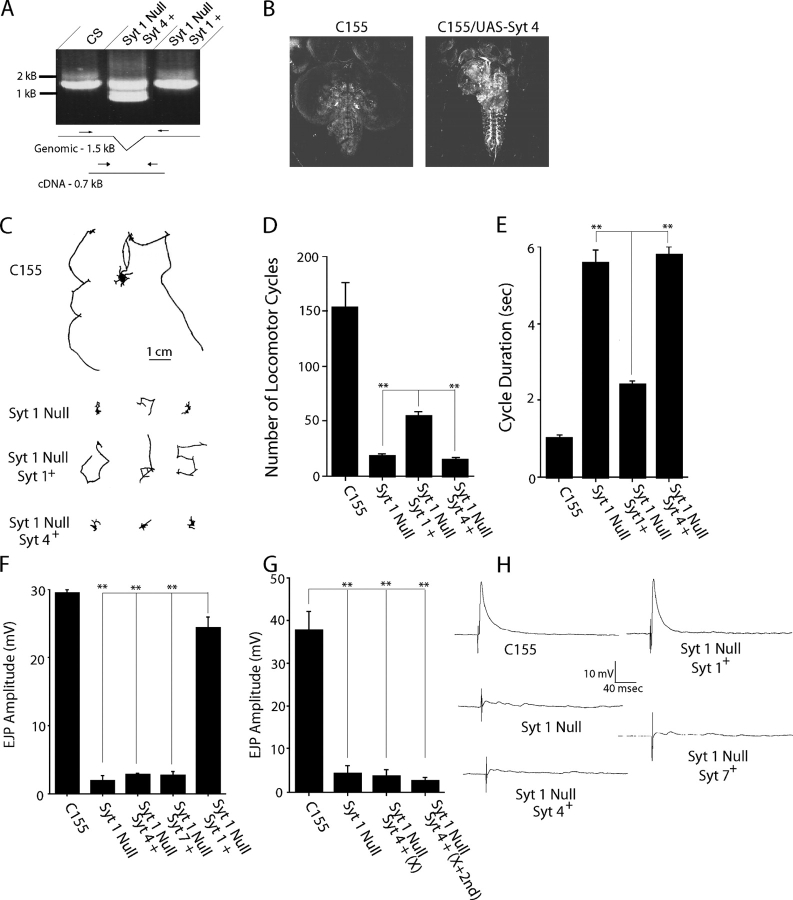
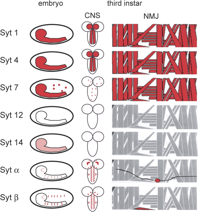
The synaptotagmin family has been implicated in calcium-dependent neurotransmitter release, although Synaptotagmin 1 is the only isoform demonstrated to control synaptic vesicle fusion. Here, we report the characterization of the six remaining synaptotagmin isoforms encoded in the Drosophila genome, including homologues of mammalian Synaptotagmins 4, 7, 12, and 14. Like Synaptotagmin 1, Synaptotagmin 4 is ubiquitously present at synapses, but localizes to the postsynaptic compartment. The remaining isoforms were not found at synapses (Synaptotagmin 7), expressed at very low levels (Synaptotagmins 12 and 14), or in subsets of putative neurosecretory cells (Synaptotagmins alpha and beta). Consistent with their distinct localizations, overexpression of Synaptotagmin 4 or 7 cannot functionally substitute for the loss of Synaptotagmin 1 in synaptic transmission. Our results indicate that synaptotagmins are differentially distributed to unique subcellular compartments. In addition, the identification of a postsynaptic synaptotagmin suggests calcium-dependent membrane-trafficking functions on both sides of the synapse.


Figures

Figure 1.
Figure 1.
Conservation of Drosophila synaptotagmins. (A) The domain structure of Drosophila synaptotagmins is shown (top). Protein sequence alignment of loops 1 and 3 reveals the conservation of the calcium-coordinating aspartic or glutamic acid residues (*) among family members (bottom). TMD, transmembrane domain. (B) Dendrogram of synaptotagmins collected from Drosophila, C. elegans, A. gambiae, F. rubripes, M. musculus, and H. sapiens (d, c, a, f, m, and h, respectively). Subfamilies are indicated by separate colors and named according to the mammalian nomenclature. Subfamilies not containing vertebrate representatives were designated with Greek letters. Subfamilies were defined by major branches in the diagram and consist of members that are more highly conserved across different species than to other members within a particular species.
Figure 2.
Figure 2.
Expression analysis of the Drosophila synaptotagmin family. (A) Developmental microarrays conducted by the Berkeley Drosophila Genome Project are shown. (top) RNA expression levels for the Drosophila synaptotagmins from 0–12 h after egg-laying as detected by Affymetrix microarray quantification. (bottom) Positive controls for developmentally expressed genes are shown. (B) Relative expression levels of the Drosophila synaptotagmins were determined by quantitative microarray analysis of either adult Canton S heads and bodies or heads only. All synaptotagmins were enriched in heads, with syt 1, syt 4, and syt 7 being the most abundant isoforms. Error bars represent SD. Embryonic expression patterns for the synaptotagmins were determined using RNA in situ hybridization on 0–22-h embryos. (C) syt 1 was abundantly expressed throughout the central and peripheral nervous systems. Similar to syt 1, syt 4 (D) and syt 7 (E) were expressed throughout the central and peripheral nervous systems. In addition to the CNS, syt 7 was observed in nonneuronal tissues. (F) syt 14 was expressed at a relatively low level in the CNS. (G) Abundant syt β signal was detected in a bilaterally symmetric population of large cells in the VNC (top left, arrowhead) and a subset of cells in the embryonic brain (bottom left). (right) High magnification view of the VNC cells is shown. Bar, 25 μm. Apart from cells in the nervous system, the syt β probe also detected several peritracheal cells (bottom left, arrowhead) present in each segment. (H) syt α expression was detected in a population of relatively small cells (left, arrowhead) in the VNC and a subset of cells in the brain (right, arrowhead).
Figure 3.
Figure 3.
Analysis of synaptotagmin subcellular compartments. (A, top) Diagram indicating the portion of the protein used to generate synaptotagmin antisera. Recombinant proteins were purified as GST fusions as described. With the exception of Syt 7, each GST fusion protein was cleaved with thrombin to remove the GST moiety. Removing GST from the Syt 7 C2 domains resulted in increased degradation, so this moiety was left attached. (top) In the bottom portion of the diagram, recombinant C2 domains of the indicated synaptotagmins were equally loaded onto a 10% polyacrylamide gel, subjected to SDS-PAGE electrophoresis, and stained with Coomassie blue. Protein preparations were then diluted 1:20 and subjected to Western analysis with the indicated polyclonal antibodies. Each antibody is specific for the synaptotagmin isoform that served as its specific antigen. (B) Post-nuclear fractions of Canton S head extracts were separated on 10–30% sucrose gradients. Isolated fractions were probed for subcellular markers by Western analysis, including antisera against Syx1A and ROP, which localize to the plasma membrane (left-most fractions). Synaptic vesicle fractions were identified using the Syt 1 and n-Synaptobrevin antibodies, cytosolic fractions were indicated by immunostaining for ROP, and endosomal fractions by staining for HRS (Lloyd et al., 2002). Syt 4 and Syt 7 were not detected in synaptic vesicle or plasma membrane fractions, but rather found near the top of the gradient. Syt α comigrated with plasma membrane markers. The last collected fraction (right-most lane) often contained contaminants from the residual membrane debris extracted from the tube sides in the final step. (C) Equilibrium density gradient fractions were probed for synaptotagmins to detect the localization of their respective compartments. Under these conditions, synaptic vesicles (Syt 1) migrate at the top of the gradient. The remaining synaptotagmins migrated to the bottom of the gradients. (D, top) Western blots of adult head extracts collected from wild-type and a syt 4 null mutant (syt4 BA1) were probed with the Syt 4 antibody. (bottom) Adult head extract isolated from either wild-type (Canton S) or animals overexpressing a syt 7 transgene and analyzed by Western analysis using the Syt 7 antibody. Extracts were collected from females (f) (C155elav -GAL4/+; +/+; UAS-syt 7/+) and males (m) (C155elav -GAL4; +/+; UAS-syt 7/+) separately. (E) Specificity of the Syt 7, Syt α, and Syt β antibodies was determined using Western analysis on Canton S adult head extract. Antibodies were incubated overnight at 4°C either with sepharose beads containing the respective GST fusion proteins or GST alone. Except for the Syt β blots, which were developed at the same time, equivalent exposure times were determined by the intensity of the Syx1A signal.
Figure 4.
Figure 4.
Characterization of Syt 4 immunoreactivity. (A) Wild-type first instar CNS immunostained with anti–Syt 4 (magenta) and a neuronal marker, anti-HRP (green). Bar, 50 μm. Syt 4–specific signal was concentrated in the neuropil of the ventral ganglion where synapses occur. (B) First instar CNS of a syt 4 deletion mutant immunostained with anti–Syt 4 (magenta) and a neuronal marker, anti-HRP (green) reveals a loss of Syt 4 immunoreactivity. Bar, 50 μm. (C) Early stage 17 embryo costained with anti–Syt 4 and anti-Fas II antibodies. Bar, 20 μm. Fas II is found in axonal tracts in the CNS, whereas Syt 4 was localized to CNS cell bodies. (D–I) Wild-type third instar neuromuscular synapses were imaged after costaining with anti-HRP and either anti–Syt 1 (D, F, and H) or anti–Syt 4 (E, G, and I). Bars, 5 μm. In contrast to Syt 1 staining, which labels synaptic vesicles localized within anti-HRP labeling of the presynaptic membrane, Syt 4 immunoreactivity is found in punctate clusters localized postsynaptically outside of anti-HRP labeling.
Figure 5.
Figure 5.
Localization of Syt 4 to postsynaptic vesicles. (A and B) Colabeling with the presynaptic membrane marker anti-HRP and anti–Syt 4. Bar, 2 μm. Confocal optical sections through a labeled third instar NMJ are shown in two axes: parallel to the body wall (X-Y; A) and perpendicular to the body wall and longitudinal (X-Z; B). Syt 4 immunoreactive clusters can be identified outside of the anti-HRP presynaptic terminal within the postsynaptic muscle (arrowheads). (C and D) Third instar neuromuscular synapses were imaged after costaining with the postsynaptic marker anti-myc antibody to detect myc-tagged GluRIIA, and either anti–Syt 1 (C) or anti–Syt 4 (D). Bar, 5 μm. Unlike Syt 1 immunoreactivity, which is found in boutons and surrounded by GluR immunoreactivity, Syt 4 concentrates at regions surrounding glutamate recep- tor clusters. (E and F) Colabeling with anti–Syt 1 and anti–Syt 4 in syt 4 null mutants overexpressing UAS-syt 4 with C155elav -GAL4. Bars: (E) 5 μm; (F) 2 μm. Although Syt 4 can be detected presynaptically when overexpressed, the Syt 4 staining is specifically excluded from Syt 1–positive synaptic vesicle domains, indicating that overexpression of Syt 4 does not cause sorting of the protein to synaptic vesicles.
Figure 6.
Figure 6.
Localization of Drosophila Syt 7. (A) Early stage 17 embryo labeled with anti–Syt 1 (magenta) and anti–Syt 7 (green) antibodies. Bar, 20 μm. Whereas Syt 1 is localized to the synaptic neuropil, Syt 7 is found within neuronal cell bodies at this stage of development. (B) Third instar NMJ stained with anti–Syt 7 antibody preabsorbed to the recombinant GST-Syt 7 fusion protein reveals no signal. Bar, 50 μm. (C) Third instar NMJ stained with anti–Syt 7 antibody preabsorbed to recombinant GST protein reveals vesicular staining throughout the muscle at sites beneath the plasma membrane. Bar, 20 μm. (D) Third instar imaginal disc stained with anti–Syt 7 antibody reveals widespread immunolocalization of Syt 7 to cell bodies. Bar, 20 μm.
Figure 7.
Figure 7.
Localization of Drosophila synaptotagmins α and β. (A) Third instar CNS stained with the anti–Syt α antibody. Bar, 50 μm. Staining was observed in the mushroom bodies and several cell bodies in the CNS. (B) Early stage 17 embryo stained with the anti–Syt α antibody. Bar, 50 μm. Specific signal was detected in two populations of cells in the CNS, one bilaterally symmetric pair (arrowheads) and another present in the midline. (C) Third instar neuromuscular preparation stained with the anti–Syt α antibody. Signal was detected in the lateral bipolar dendritic neuron (arrow). Bar, 20 μm. (D) Syt β antibody staining of third instar NMJs. Bar, 50 μm. Fluorescence image was overlaid onto the DIC image to indicate muscle positions. Syt β staining was observed only at motor terminals innervating muscle fiber 8. (E) Third instar CNS stained with the anti–Syt β antibody. Bar, 50 μm. The antibody labels several cell bodies throughout the CNS. The immunolocalization was distinct from anti–Syt α staining, suggesting unique subpopulations of neurons expressing each isoform. (F) Staining of peritracheal cells located at tracheal branchpoints in late stage embryos is also observed with the anti–Syt β antibody, consistent with in situ labeling of the same cells. Bar, 20 μm.
Figure 8.
Figure 8.
Syt 4 and Syt 7 cannot rescue release defects in syt 1 mutants. (A) PCR confirmation of the syt 4 transgene in animals used for rescue experiments was obtained by priming across a small intron, revealing a larger 1.5-kB band from the native genomic locus, and a 0.7-kB band specifically from animals containing the UAS-syt 4 cDNA lacking the intron. (B) Immunostaining with anti–Syt 4 antibodies from control and C155elav -GAL4/UAS-syt 4; syt AD4/Df(2L)N13 lines. The confocal settings were identical between the two pictures, and the signal intensity was set to a low level to highlight the strong up-regulation of Syt 4 in the third instar CNS of syt 1 null animals containing UAS-syt 4 and the C155elav -GAL4 driver. (C) Traces of the crawling pattern of control, syt 1 null mutants, and rescued lines containing UAS-syt 1 or UAS-syt 4 are shown for a 4-min imaging period. Quantification of the number of locomotor cycles during 4 min (D) and the cycle duration (E) are shown. Error bars are SEM. Similar results were observed when UAS-syt 1 and UAS-syt 4 were driven with a third chromosome elav-GAL4 driver (not depicted). The number of animals examined were as follows (number for locomotor cycle number, number for locomotor cycle duration): C155elav -GAL4, n = 5, 5; syt AD4/Df(2L)N13, n = 17, 7; C155elav -GAL4/UAS-syt 1; syt AD4/Df(2L)N13, n = 15, 5; and C155elav -GAL4/UAS-syt 4; syt AD4/Df(2L)N13, n = 8, 7. (F) Mean evoked EJP amplitudes (± SEM) recorded in 1.5 mM extracellular calcium for the indicated genotypes. In contrast to the rescue observed with syt 1 transgenic expression, Syt 4 and Syt 7 had no effect on neurotransmission in the syt 1 null mutant. Average muscle resting potentials ± SD were unchanged between the genotypes and were as follows: C155elav -GAL4, 59.3 ± 3.9 (n = 26); syt AD4/Df(2L)N13, 61.1 ± 5.2 (n = 17); C155elav -GAL4/UAS-syt 1; syt AD4/Df(2L)N13, 63.7 ± 3.6 (n = 10); C155elav -GAL4/+; syt AD4/Df(2L)N13; UAS-syt 7/+, 56.1 ± 3.4 (n = 27); and C155elav -GAL4/UAS-syt 4; syt AD4/Df(2L)N13, 61.5 ± 4.4 (n = 16). In 10% of animals containing the syt 7 transgene, a small degree of rescue was observed, with evoked responses averaging ∼30% of the response observed in syt 1 rescued control animals. The other 23 animals showed no rescue, and the results shown are pooled data from both sets of syt 7 animals. No case of rescue was observed in UAS-syt 4 overexpression experiments. (G) Mean evoked EJP amplitudes (± SEM) recorded in 5.0 mM extracellular calcium for the indicated genotypes. Average muscle resting potentials ± SD were unchanged between the genotypes and were as follows: C155elav-GAL4, 62.1 ± 4.2 (n = 25); syt AD4/Df(2L)N13, 59.4 ± 3.8 (n = 18); C155elav -GAL4/UAS-syt 4; syt AD4/Df(2L)N13, 57.2 ± 3.8 (n = 26); and C155elav -GAL4/UAS-syt 4; elav-GAL4, syt AD4/Df(2L)N13, 59.3 ± 4.0 (n = 5). (H) Representative traces of evoked responses at 1.5 mM extracellular calcium for the indicated genotypes. In contrast to the fast release observed in control and Syt 1 rescued animals, Syt 4 and Syt 7 rescued animals and the syt 1 null mutant showed only slow EJPs, reflecting asynchronous synaptic transmission. Statistical significance was determined by t test; **, P < 0.001.
Figure 9.
Figure 9.
Summary of the expression pattern of the Drosophila synaptotagmin family. The results from embryonic in situ experiments are shown in the left panel, whereas the two right panels highlight protein expression in the third instar larval CNS and periphery. The muscles labeled red indicate NMJs where presynaptic localization of Syt 1, Syt α, or Syt β occurs, postsynaptic localization of Syt 4, and general sarcoplasmic localization of Syt 7.

References

    1. Adams, M.D., S.E. Celniker, R.A. Holt, C.A. Evans, J.D. Gocayne, P.G. Amanatides, S.E. Scherer, P.W. Li, R.A. Hoskins, R.F. Galle, et al. 2000. The genome sequence of Drosophila melanogaster. Science. 287:2185–2195. - PubMed
    1. Adolfsen, B., and J.T. Littleton. 2001. Genetic and molecular analysis of the synaptotagmin family. Cell. Mol. Life Sci. 58:393–402. - PMC - PubMed
    1. Aparicio, S., J. Chapman, E. Stupka, N. Putnam, J.M. Chia, P. Dehal, A. Christoffels, S. Rash, S. Hoon, A. Smit, et al. 2002. Whole-genome shotgun assembly and analysis of the genome of Fugu rubripes. Science. 297:1301–1310. - PubMed
    1. Butz, S., R. Fernandez-Chacon, F. Schmitz, R. Jahn, and T.C. Südhof. 1999. The subcellular localizations of atypical synaptotagmins III and VI. Synaptotagmin III is enriched in synapses and synaptic plasma membranes but not in synaptic vesicles. J. Biol. Chem. 274:18290–18296. - PubMed
    1. Cantera, R., and D.R. Nassel. 1992. Segmental peptidergic innervation of abdominal targets in larval and adult dipteran insects revealed with an antiserum against leucokinin I. Cell Tissue Res. 269:459–471. - PubMed

Publication types