. 2004 Jul 19;200(2):159-68.
doi: 10.1084/jem.20031975.

A role for thymic stromal lymphopoietin in CD4(+) T cell development

Affiliations

A role for thymic stromal lymphopoietin in CD4(+) T cell development

Amin Al-Shami et al. J Exp Med. .

Abstract

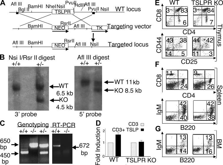
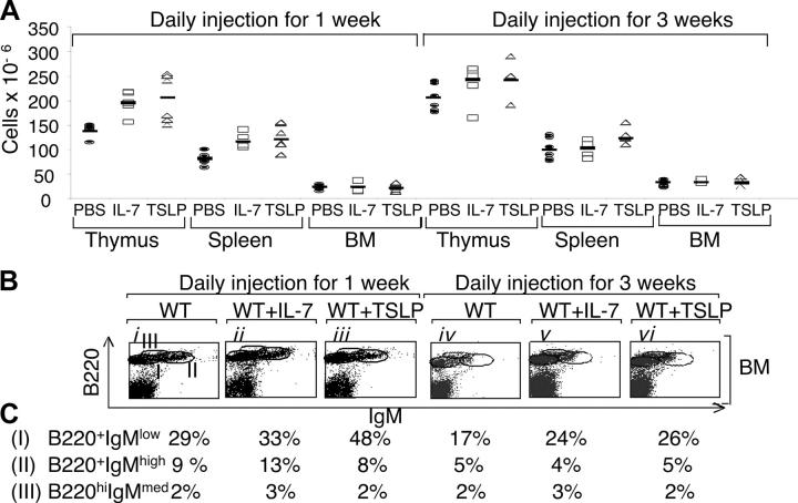
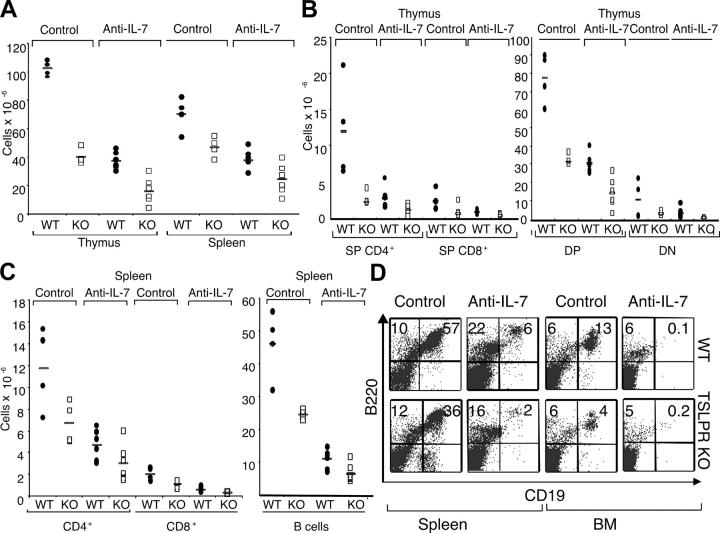
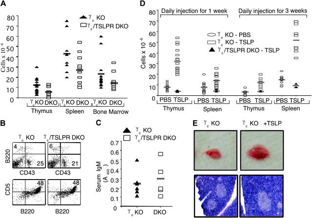
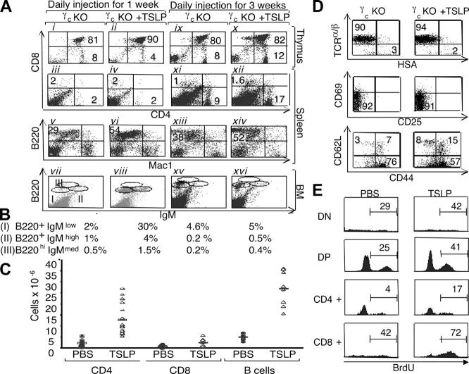
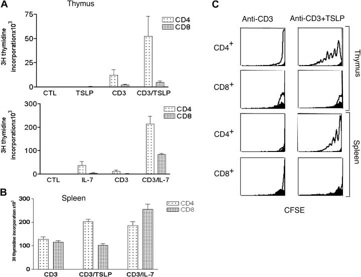
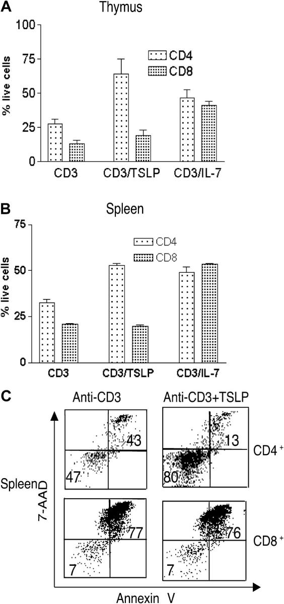
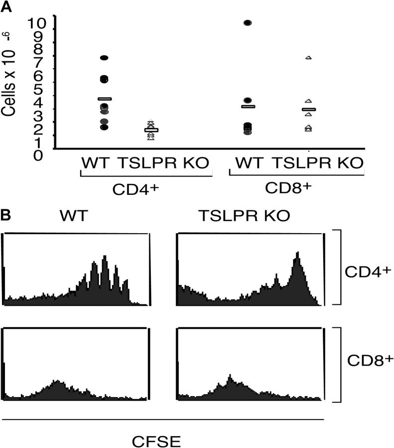
Thymic stromal lymphopoietin (TSLP) signals via a receptor comprising the interleukin (IL)-7 receptor alpha chain and a distinctive subunit, TSLP receptor (TSLPR), which is most related to the common cytokine receptor gamma chain, gamma(c). We have generated TSLPR knockout (KO) mice and found that although these mice had normal lymphocyte numbers, gamma(c)/TSLPR double KO mice had a greater lymphoid defect than gamma(c) KO mice. This indicates that TSLP contributes to lymphoid development and accounts for some of the residual lymphoid development in gamma(c) KO mice and presumably in patients with X-linked severe combined immunodeficiency. Injection of TSLP into gamma(c) KO mice induced the expansion of T and B cells. Moreover, sublethally irradiated TSLPR KO mice showed weaker recovery of lymphocyte populations than wild-type (WT) littermates, even when neutralizing anti-IL-7 antibodies were injected. Interestingly, TSLP preferentially stimulated the proliferation and survival of CD4(+) single positive thymocytes and peripheral T cells in vitro. Additionally, CD4(+) T cells from TSLPR KO mice expanded less efficiently than WT CD4(+) T cells in irradiated hosts, and TSLP preferentially expanded CD4(+) T cells both in vitro and in vivo. Thus, as compared with other known cytokines, TSLP is distinctive in exhibiting a lineage preference for the expansion and survival of CD4(+) T cells.


Figures

Figure 1.
Figure 1.
Normal immunological development in TSLPR KO mice. (A) Schematic of the TSLPR targeting strategy. The 6-kb BglII to Nhe I 5′ and 3-kb Pvu II 3′ flanking regions of the TSLPR gene were cloned 5′ and 3′ to the Neo gene. The targeting vector was linearized, electroporated into ES cells, and transfected clones were screened using 5 and 3′ probes (squares). (B) Southern hybridization of the ES clones. The restriction enzymes and fragment sizes are indicated (+/+, WT; +/−, heterozygote). (C) Genotyping of the mice was performed as described in Materials and Methods (left). RT-PCR was performed using TSLPR internal primers to detect the TSLPR mRNA transcript thymus (right). White lines indicate that intervening lanes have been spliced out. (D) TSLP significantly increased anti-CD3ɛ–induced proliferation of WT splenocytes (P < 0.001), but not of TSLPR KO splenocytes. Results are expressed as mean fold induction ± SEM (n = 14). (E–G) Flow cytometric analysis showing no difference in the CD4/CD8 profile of the total thymus (E, top), the CD25/CD44 profile of DN thymocytes (E, bottom), or in the spleen (F) and BM (G) of WT and TSLPR-deficient mice. Antibodies for the CD4+ and CD8+ surface markers were used to evaluate the T cell populations in the thymus (E) and spleen (F, top), whereas antibodies for B220+ and surface IgM+ were used to evaluate B cells populations in the spleen (F, bottom) and BM (G).
Figure 2.
Figure 2.
TSLP can expand lymphocyte populations in WT mice. (A) The absolute number of thymocytes, splenocytes, and BM cells in WT mice injected daily with PBS, IL-7, or TSLP for 1 and 3 wk. 1 wk of treatment with TSLP and IL-7 increased thymic cellularity (means ± SEM of 204 ± 46 × 106 and 195 ± 22 × 106 [P = 0.01 and 0.001, respectively] vs. 139 ± 13 × 106 for the PBS-treated mice). 1 wk of treatment with TSLP and IL-7 also increased splenic cellularity (means ± SEM of 126 ± 23 × 106 and 118 ± 13 × 106 [P = 0.004 and 0.002, respectively] vs. 80 ± 12 × 106 for the PBS-treated mice). No significant difference was observed in the BM. After 3 wk of treatment with TSLP or IL-7, the changes in cellularity of thymus, spleen, and BM were not significant. (B) Flow cytometric analysis of the BM 1 and 3 wk after injection of WT mice with PBS, IL-7, or TSLP. (C) Percentages of populations shown in B.
Figure 3.
Figure 3.
TSLP is critical for optimal lymphopoiesis in the presence and absence of IL-7. WT and TSLPR KO mice were sublethally irradiated and injected three times a week for 4 wk with 1 mg of control or M25 anti–IL-7 mAbs. (A) TSLPR KO treated with control or anti–IL-7 mAbs displayed lower thymic cellularity (P < 0.001 for both mAbs) and splenic cellularity (P = 0.03 for anti–IL-7 and P = 0.02 for control mAb) when compared with WT littermates. (B) The absolute numbers of thymic subpopulations, except for DN cells, were decreased in TSLPR KO mice compared with WT mice (P < 0.05). (C) The absolute numbers of CD4+ and CD8+ T and B cells in the spleens of irradiated animals. Mice lacking TSLPR had fewer lymphocytes than WT mice, when experiments were performed with either control or anti–IL-7 mAbs (P < 0.05). (D) B220 versus CD19 flow cytometric analysis of the spleen and BM from WT (top) and TSLPR KO (bottom) mice.
Figure 4.
Figure 4.
Comparison of the lymphoid development in γc KO mice versus γc/TSLPR DKO mice. (A) γc/TSLPR DKO mice had lower thymic, spleen, and BM cellularities than γc KO mice (thymus: mean ± SEM of 12.3 ± 6.7 × 10⁶ for γc KO mice vs. 5.7 ± 4.5 × 10⁶ for γc/TSLPR DKO mice [P = 0.009]; spleen: mean ± SEM of 43 ± 17.7 × 10⁶ for γc KO mice vs. 26.8 ± 13.7 × 10⁶ for γc/TSLPR DKO mice [P = 0.02]; BM: mean ± SEM of 23.4 ± 15.5 × 10⁶ for γc KO mice vs. 14.3 ± 7.6 × 10⁶ for γc/TSLPR DKO mice [P = 0.047]). All mice were age matched and no sex-related differences were noted. (B) Flow cytometric analysis of BM (top, B220 vs. CD43) and peritoneal cavity lymphocytes (bottom, B220 vs. CD5). (C) Similar levels of IgM in the serum of γc KO and γc/TSLPR DKO mice. (D) Injection of 0.5 μg/day of TSLP (open rectangles) enhances lymphoid cellularity in γc KO mice. Treatment for 1 and 3 wk showed an enhanced cellularity in thymus (mean ± SEM of 7.8 ± 2.9 × 10⁶ for PBS-injected mice vs. 32 ± 10 × 106 for TSLP-injected mice [P < 0.0001] after 1 wk of treatment and a mean ± SEM of 3.5 ± 2 × 10⁶ for PBS-injected mice vs. 12.2 ± 4 × 10⁶ for TSLP-injected mice [P = 0.001] after 3 wk) and spleen (mean ± SEM of 8 ± 4 × 10⁶ for PBS-injected mice vs. 14 ± 5.4 × 10⁶ for TSLP-injected mice [P = 0.003] after 1 wk of treatment and a mean ± SEM of 15 ± 4 × 10⁶ for PBS-injected mice vs. 52 ± 12 × 106 for TSLP-injected mice [P < 0.0001] after 3 wk). No change was seen in the BM. All mice were age matched and no sex-related differences were noted. γc/TSLPR DKO mice did not respond to TSLP injections (closed triangles). (E) Photograph indicating thymic size in γc KO mice after wk of treatment with PBS or TSLP (top) and a histological analysis of these tissues by hematoxylin and eosin staining (bottom).
Figure 5.
Figure 5.
TSLP can increase lymphoid subpopulations in γc-deficient mice. (A) Flow cytometric analysis of γc KO thymus, spleen, and BM 1 and 3 wk after injection of PBS or TSLP. (B) B cell populations in the BM from A (vii, viii, xv, and xvi). (C) TSLP injections induced an increase in CD4+ T cells (mean ± SEM of 2.36 ± 1.48 × 10⁶ for control mice vs. 12.7 ± 6.7 × 10⁶ for TSLP-treated mice [P < 0.0001], for a 5.4-fold increase), CD8+ T cells (mean ± SEM of 0.63 ± 0.43 × 10⁶ for control mice vs. 2.4 ± 1.7 × 10⁶ for TSLP-treated mice [P = 0.01], for a 3.8-fold increase), as well as B cells (mean ± SEM of 4.6 ± 1.2 × 10⁶ for control mice vs. 27 ± 7 × 10⁶ for TSLP-treated mice [P < 0.0001], for a 5.9-fold increase) in the spleen of γc KO mice 3 wk after injection. (D) Flow cytometric analysis of splenic CD4+ T cells in γc mice treated with PBS or TSLP for 3 wk. TSLP increased the absolute numbers of CD44high CD62Llow (mean ± SEM of 2.3 ± 1.2 × 10⁶ for control γc mice vs. 11.3 ± 4.106 for TSLP-treated mice [P = 0.0004], for 4.9-fold increase), CD44high CD62Lhigh (mean ± SEM of 0.4 ± 0.2 × 10⁶ for control γc mice vs. 3.2 ± 1.1 × 10⁶ for TSLP-treated mice [P = 0.0003], for an eightfold increase), and CD44low CD62Lhigh (mean ± SEM of 0.08 ± 0.06 × 10⁶ for control γc mice vs. 0.9 ± 0.16 × 10⁶ for TSLP-treated mice [P < 0.0001], for an 11-fold increase). (E) γc KO mice were treated with PBS or TSLP for 1 wk and injected with BrdU 10 and 16 h before being killed. BrdU incorporation was measured by intracellular staining using PE-labeled BrdU of the thymocytes subpopulations. The number indicates the percent of BrdU+ cells within the gated region.
Figure 6.
Figure 6.
TSLP preferentially expands CD4+ T cells. In A and B, cells were treated with medium, 100 ng/ml IL-7, or 100 ng/ml TSLP, and/or 2 μg/ml anti-CD3ɛ antibodies. (A) In vitro proliferation of purified CD4+ and CD8+ SP thymocytes from WT mice. Results are expressed as mean ± SEM for four experiments. TSLP increased anti-CD3ɛ–induced proliferation of CD4+ SP cells (P = 0.02), but did not significantly affect CD8+ SP expansion to anti-CD3ɛ (P = 0.07). IL-7 significantly enhanced anti-CD3ɛ–induced expansion of both CD4+ and CD8+ SP thymocytes (P < 0.0001 for both). (B) In vitro proliferation of purified CD4+ and CD8+ splenocytes (prepared by positive selection) treated as described above. TSLP significantly increased anti-CD3ɛ–induced proliferation of mature CD4+ T cells (P = 0.0002), but not of CD8+ T cells (P = 0.1). IL-7 significantly enhanced anti-CD3ɛ–induced expansion of both CD4+ and CD8+ mature T cells (P = 0.008 and 0.0005, respectively). (C) CD4+ thymocytes and splenic T cells were labeled with CFSE and cultured for 1 wk with anti-CD3ɛ with or without TSLP, and cells were analyzed by flow cytometry. As evaluated by decreased CFSE staining, TSLP increased anti-CD3ɛ–induced proliferation of CD4+ but not CD8+ T cells. WT corresponds to the “open” curve, whereas TSLPR KO mice are shown in solid black.
Figure 7.
Figure 7.
TSLP promotes survival of CD4+ T cells. (A) In vitro survival of purified CD4+ and CD8+ SP thymocytes from WT mice. The percent of viable cells (mean ± SEM for four experiments) was determined after 1 wk by trypan blue exclusion. TSLP increased anti-CD3ɛ–induced survival of CD4+ cells (P = 0.02), but did not significantly affect CD8+ survival (P = 0.24). IL-7 significantly enhanced anti-CD3ɛ–induced survival of both CD4+ and CD8+ thymocytes (P = 0.03 and P < 0.0001, respectively). (B) In vitro survival assay of purified CD4+ and CD8+ splenic T cells from WT mice. The percent of viable cells was determined by trypan blue exclusion. Results are expressed as mean ± SEM for five experiments. TSLP significantly increased anti-CD3ɛ–induced survival of CD4+ (P < 0.0001) but not CD8+ T cells (P = 0.21). IL-7 significantly enhanced anti-CD3ɛ–induced survival of both CD4+ and CD8+ T cells (P < 0.001 and P < 0.0001, respectively). (C) Purified CD4+ and CD8+ splenocytes cultured as indicated were stained with annexin V and 7-AAD.
Figure 8.
Figure 8.
TSLP mediates efficient expansion of CD4+ T cells. CD4+ T cells were isolated from WT or TSLPR KO mice and labeled with CFSE before being injected into irradiated γc KO mice. (A) After 1 wk, TSLPR KO CD4+ T cells expanded less than CD4+ T cells from WT mice (P = 0.008). CD8+ T cells from WT or TSLPR mice expanded to a similar degree. (B) Examination of the CFSE dilution on day 3 by flow cytometry revealed that WT CD4+ T cells were expanding more rapidly than TSLPR KO CD4+ T cells. No differences were observed for CD8+ T cells.

References

    1. Leonard, W.J. 2003. Type I Cytokines and Interferons and Their Receptors. Fundamental Immunology. W.E. Paul, editor. Lippincott-Williams and Williams, Philadelphia. 701–747.
    1. Peschon, J.J., P.J. Morrissey, K.H. Grabstein, F.J. Ramsdell, E. Maraskovsky, B.C. Gliniak, L.S. Park, S.F. Ziegler, D.E. Williams, C.B. Ware, et al. 1994. Early lymphocyte expansion is severely impaired in interleukin 7 receptor–deficient mice. J. Exp. Med. 180:1955–1960. - PMC - PubMed
    1. von Freeden-Jeffry, U., P. Vieira, L.A. Lucian, T. McNeil, S.E. Burdach, and R. Murray. 1995. Lymphopenia in interleukin (IL)-7 gene-deleted mice identifies IL-7 as a nonredundant cytokine. J. Exp. Med. 181:1519–1526. - PMC - PubMed
    1. Puel, A., S.F. Ziegler, R.H. Buckley, and W.J. Leonard. 1998. Defective IL7R expression in T(−)B(+)NK(+) severe combined immunodeficiency. Nat. Genet. 20:394–397. - PubMed
    1. Friend, S.L., S. Hosier, A. Nelson, D. Foxworthe, D.E. Williams, and A. Farr. 1994. A thymic stromal cell line supports in vitro development of surface IgM+ B cells and produces a novel growth factor affecting B and T lineage cells. Exp. Hematol. 22:321–328. - PubMed

MeSH terms