. 2004 Jul 19;200(2):261-6.
doi: 10.1084/jem.20040526.

SH2D1A regulates T-dependent humoral autoimmunity

Affiliations

SH2D1A regulates T-dependent humoral autoimmunity

Jonathan D Hron et al. J Exp Med. .

Abstract

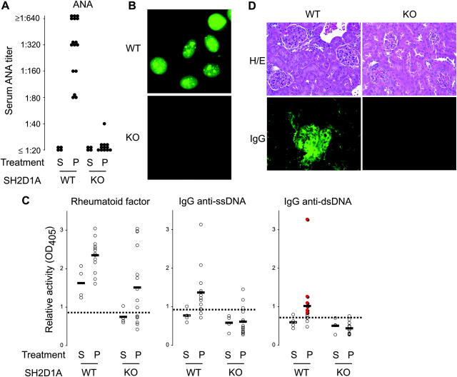
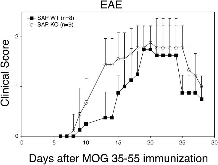
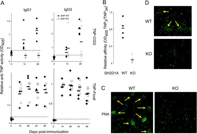
The signaling lymphocytic activation molecule (SLAM)/CD150 family includes a family of chromosome 1-encoded cell surface molecules with costimulatory functions mediated in part by the adaptor protein SH2D1A (SLAM-associated protein, SAP). Deficiency in SH2D1A protects mice from an experimental model of lupus, including the development of hypergammaglobulinemia, autoantibodies including anti-double stranded DNA, and renal disease. This protection did not reflect grossly defective T or B cell function per se because SH2D1A-deficient mice were susceptible to experimental autoimmune encephalomyelitis, a T cell-dependent disease, and they were capable of mounting normal T-independent antigen-specific immunoglobulin responses. Instead, T-dependent antibody responses were impaired in SH2D1A-deficient mice, reflecting defective germinal center formation. These findings demonstrate a specific role for the SLAM-SH2D1A system in the regulation of T-dependent humoral immune responses, implicating members of the CD150-SH2D1A family as targets in the pathogenesis and therapy of antibody-mediated autoimmune and allergic diseases.


Figures

Figure 1.
Figure 1.
SH2D1A-deficient mice are protected from experimentally induced lupus. 6–8-wk-old SH2D1A-deficient (KO) or -sufficient (WT) mice were injected intraperitoneally with 0.5 ml pristane (P) or PBS (S, saline). (A and B) 5 mo later, sera were assayed and titered for the presence of ANAs (ANA). The representative ANA fluorescence pattern of a WT versus KO serum assayed at a 1:40 dilution is shown in B. (C) Rheumatoid factor and anti-ssDNA and anti-dsDNA titers were determined by ELISA. Specimens positive by Crithidia lucillae immunofluorescence are indicated in the anti-dsDNA graph in red. n = 4, 16, 4, and 18 for WT − P, WT − S, KO − P, and KO − S, respectively. Dotted lines indicate thresholds for positivity, as determined by three standard deviations above the mean of nonautoimmune BALB/c mice. (D) Representative renal histology (H/E) and glomerular IgG deposition (IgG) of WT versus KO animals 15 wk after treatment with pristane. Note the development of mesangila wall thickening and glomerular hypercellularity associated with IgG deposition in WT, but not KO, kidneys.
Figure 2.
Figure 2.
SH2D1A-deficient mice are susceptible to experimental autoimmune EAE. EAE was induced in 5–6-wk-old SH2D1A-deficient (KO) or -sufficient (WT) mice using the MOG38-50 peptide. Clinical score was determined daily on each animal. Data points indicate means ± standard deviations.
Figure 3.
Figure 3.
Defective T-dependent, but not T-independent, humoral immunity in the absence of SH2D1A. 6-wk-old SH2D1A-deficient (KO) or -sufficient (WT) mice were immunized intraperitoneally with 50 μg TNP-CGG (T-dependent) or TNP-Ficoll (T-independent). (A) At the times indicated, sera were assessed for relative antihapten (TNP) antibody activity by ELISA using TNP(34)-BSA and IgG isotype-specific reagents. (B) Relative anti-TNP affinities of sera from day 35 TNP-CGG–immunized animals were evaluated by taking the ratio of activities obtained per serum against TNP(3)-BSA and TNP(34)-BSA (n = 5 in each group). (C and D) Germinal centers (yellow arrows) were identified in frozen spleen sections via staining with FITC-conjugated PNA on (C) day 28 TNP-CGG–immunized animals and (D) week 15 pristane-treated animals. Specimens shown in C and D are representative of at least five animals of each genotype. Data shown represent at least two independently performed trials.

References

    1. Engel, P., M.J. Eck, and C. Terhorst. 2003. The SAP and SLAM families in immune responses and X-linked lymphoproliferative disease. Nat. Rev. Immunol. 3:813–821. - PubMed
    1. Cocks, B.G., C.C. Chang, J.M. Carballido, H. Yssel, J.E. de Vries, and G. Aversa. 1995. A novel receptor involved in T-cell activation. Nature. 376:260–263. - PubMed
    1. Lee, K.-M., M.E. McNerney, S.E. Stepp, P.A. Mathew, J.D. Schatzle, M. Bennett, and V. Kumar. 2004. 2B4 acts as a non–major histocompatibility complex binding inhibitory receptor on mouse natural killer cells. J. Exp. Med. 199:1245–1254. - PMC - PubMed
    1. Wang, N., A. Satoskar, W. Faubion, D. Howie, S. Okamoto, S. Feske, C. Gullo, K. Clarke, M.R. Sosa, A.H. Sharpe, et al. 2004. The cell surface receptor SLAM controls T cell and macrophage functions. J. Exp. Med. 199:1255–1264. - PMC - PubMed
    1. Czar, M.J., E.N. Kersh, L.A. Mijares, G. Lanier, J. Lewis, G. Yap, A. Chen, A. Sher, C.S. Duckett, R. Ahmed, et al. 2001. Altered lymphocyte responses and cytokine production in mice deficient in the X-linked lymphoproliferative disease gene SH2D1A/DSHP/SAP. Proc. Natl. Acad. Sci. USA. 98:7449–7454. - PMC - PubMed

Publication types

MeSH terms

Substances